Art Conservation Microscope Solutions
Analyzing, restoring, conserving and documenting artistic works or cultural artifacts require complex technical skills and the use of state-of-the-art imaging solutions, particularly contact-free, nondestructive methods. Combined with digital camera technology and image analysis software, Leica Microsystems’ art conservation microscope solutions are designed for the different fields of activity of restorers, art historians, archaeologists and experts in conservation workshops and museums. All microscopy solutions optimize detail-accurate work on the specimen, microscopic analysis of structures and materials as well as documentation.
Please contact us if you would like to have personal expert advice on our microscopy solutions for Art Conservation

So that you never miss a detail
Leica Microsystems provides a range of solutions for the different fields of activity of restorers, art historians, archaeologists and experts from related fields. All optimize detail-accurate work on the specimen, microscopic analysis of structures and materials as well as documentation.
Expertise for experts
Microscope solutions specially adapted to the specific requirements, combined with digital camera technology and image analysis software from Leica Microsystems, are among the professional tools used today by many experts in conservation workshops and museums.
To give you an overview of the various methods and technologies for each application area, we have listed the products that support you in your work with valuable treasures of art and irreplaceable originals.
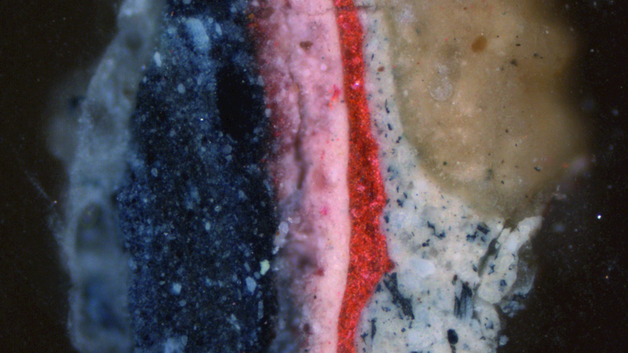
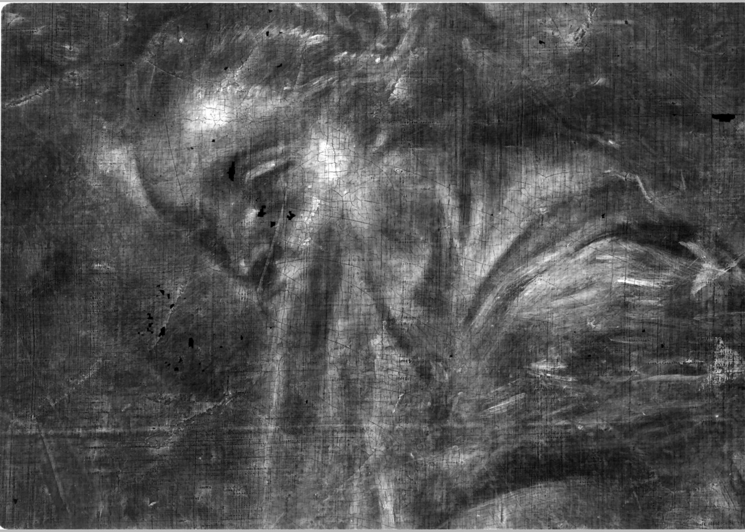
Pigment Analysis
When you examine paint layers and paint pigments, you need high-resolution, high-performance microscopes with brilliant, cool illumination. Leica Microsystems provide powerful microscopy solutions with fluorescence illumination options combined with easy-to-use analysis software for optimal investigation, analysis and documentation.
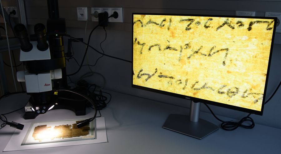
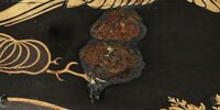
The Graz Mummy Book
‘The Graz Mummy Book’ , UBG Ms I 1946, was discovered at Graz University Library as being the oldest handwritten document in book form. It had been excavated at El-Hibeh, Egypt, and dates from 260 BCE.
The document is at least 400 years older than other existing codices, making it the earliest direct precursor of the codex form. In the image, it is being examined with an M80 stereo microscope from Leica Microsystems.
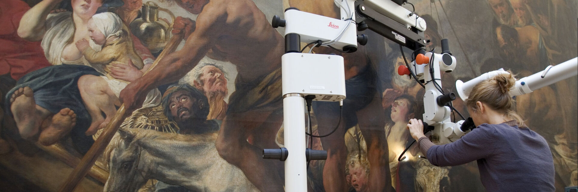
Paintings
Detail-accurate cleaning and restoration of paintings involve the use of precise digital and stereomicroscopes customized for a variety of painting types and sizes. Leica’s microscopes for paint cleaning and restoration provide variable zoom positions which enable you to work at low magnifications and analyze the tiniest details. Floor stand options are available for larger paintings.
materials
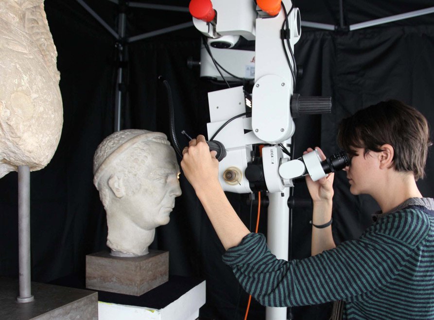
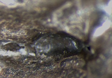
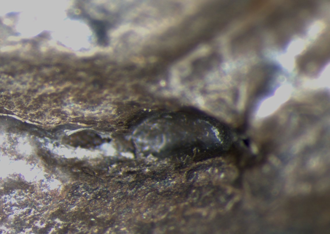
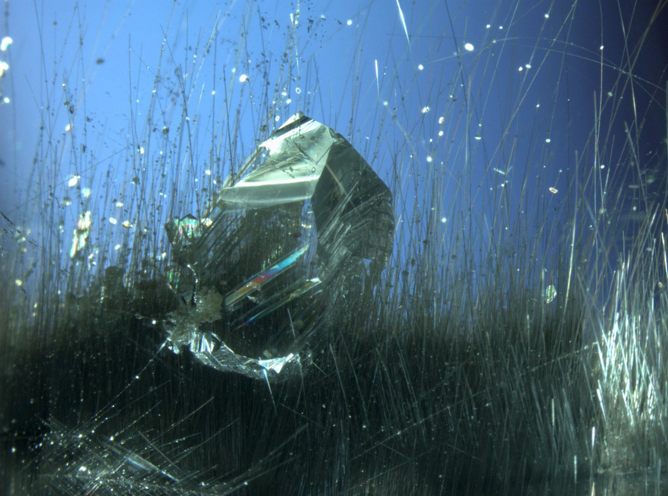
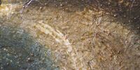
Minerals & Fossils
Mineral and fossil conservation requires detailed image processing and analysis of through high-performance stereo and research microscopes. Whether you need to view microstructures in 3D or you need to analyze thin sections, Leica Microsystems offers versatile microscopy solutions to meet your specific application needs.
Paper, Books & Photos
Reveal and analyze unique details of printing and finishing techniques, manuscripts or retouches and view the finest paper structures in 3D with Leica’s range of microscopy solutions. These innovative digital and stereomicroscopes combine maximum resolution and the greatest depth of field to support the acquiring, storing, annotating and documenting you need to research and restore these precious items of our heritage.
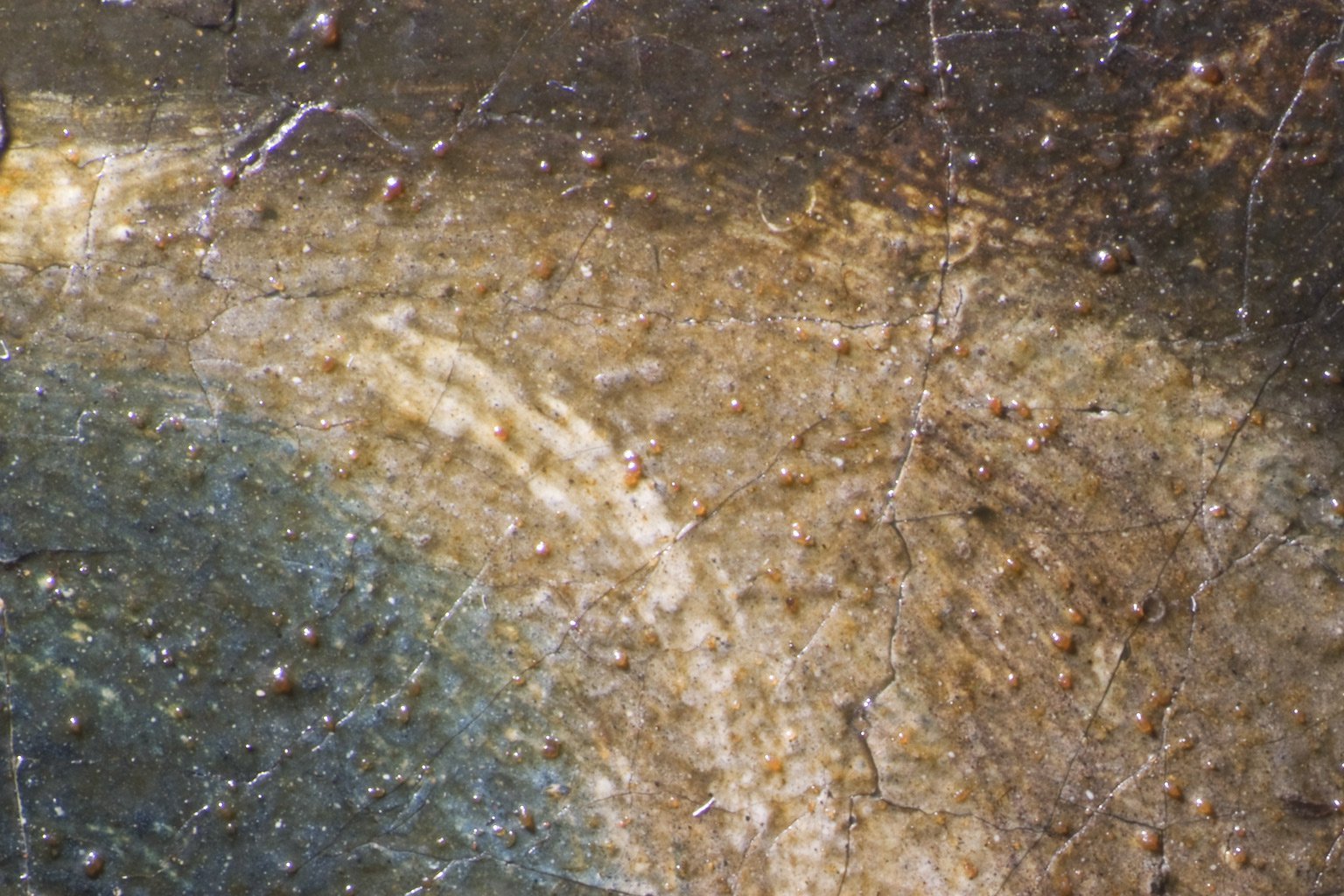
Glass, Ceramics & Stone
When you examine, clear or reconstruct glass, ceramics or stone, you need customized, high-resolution microscope solutions that enable you to analyze objects of different sizes and shapes. Leica Microsystems provides a variety of stereo and research microscopes with different illumination methods and portable stand systems tailored to your needs.
Metal
Examine signs of corrosion, dirt deposits, treatment or processing techniques for metal objects or analyzing the metal structure and determining alloy components with Leica Microsystems' individualized and affordable system solutions. Featured stereo and research microscopes are combined with digital cameras and easy-to-use 3D software to support your analysis, cleaning and documentation of metal objects.
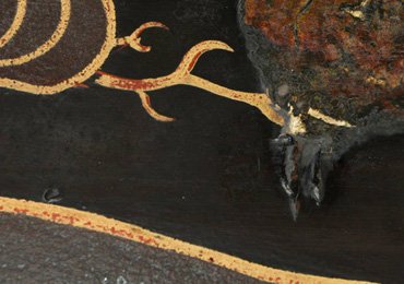
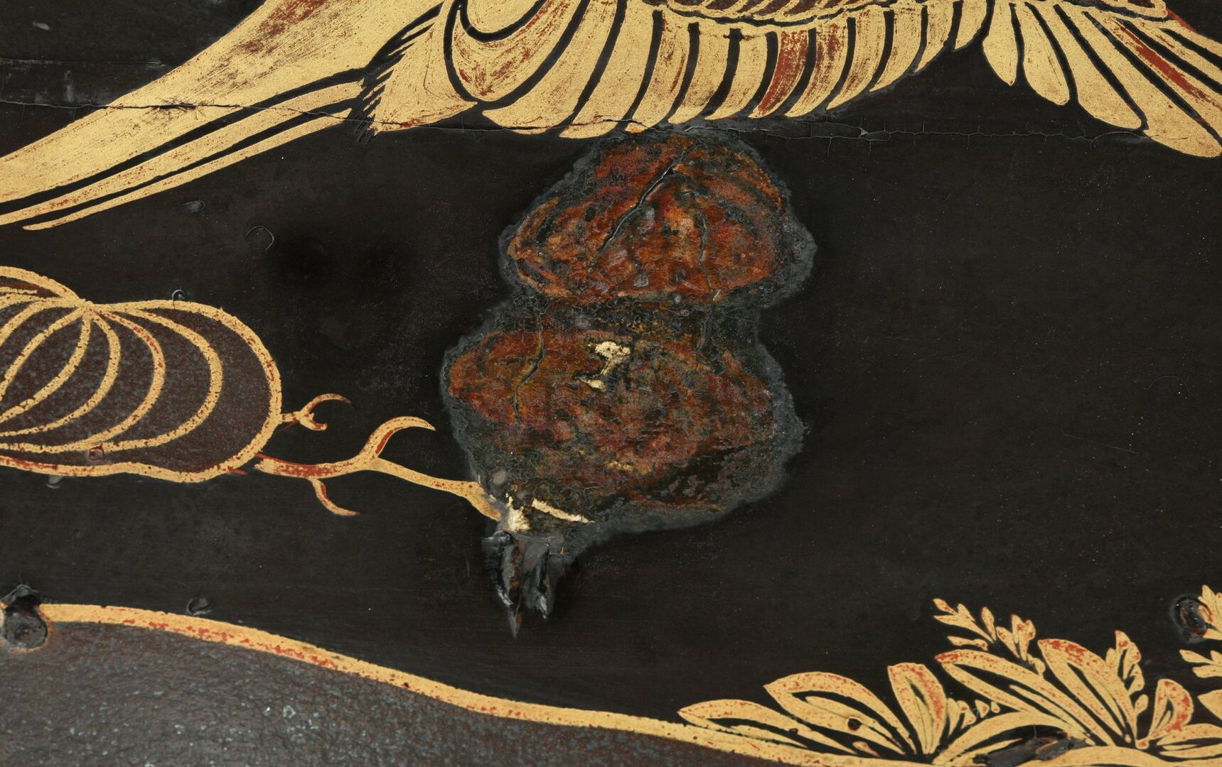
Wood
Clean and restore painted or varnished wood objects and determine the structure and components of the applied layers with Leica's stereo and research microscope solutions. These flexible solutions are combined with table or floor stands, digital cameras and easy-to-use analysis software to support your wood conservation work.


. With DIC users are able to visualize small height differences on the wafer surface more easily.")

 at the edge of a battery electrode acquired with a DVM6 digital microscope.")


