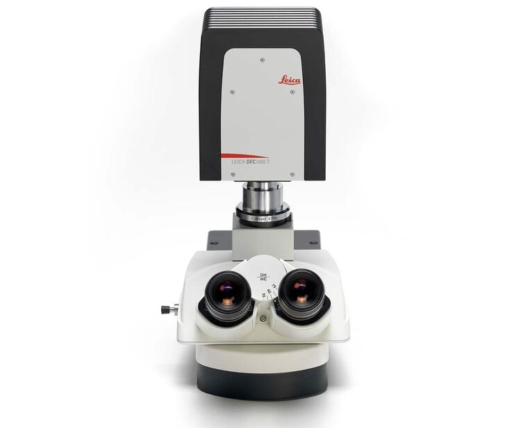
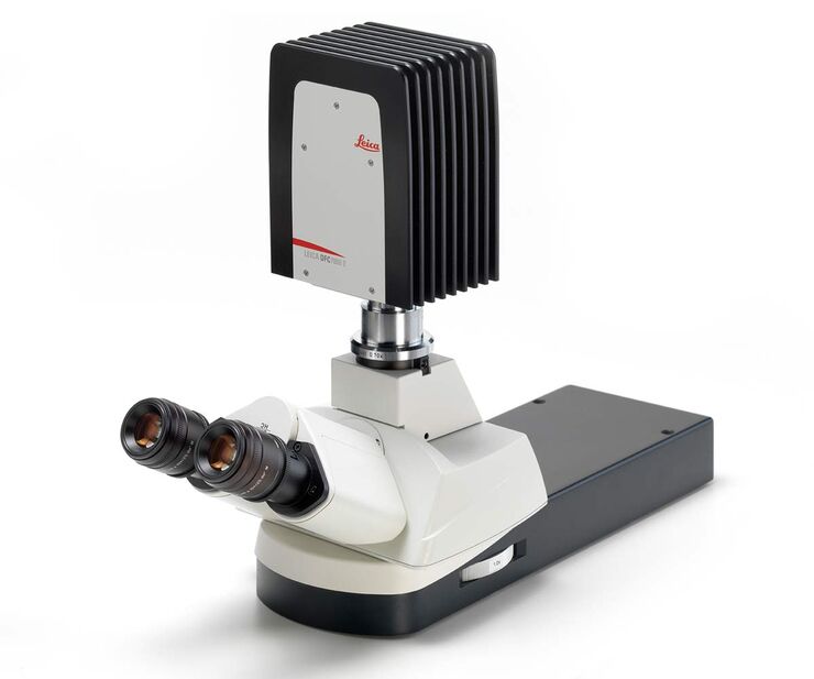
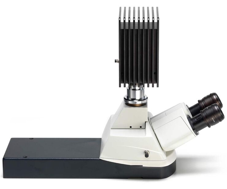
DFC7000 T and DFC7000 GT Microscope Cameras
Pro Class for Fluorescence!
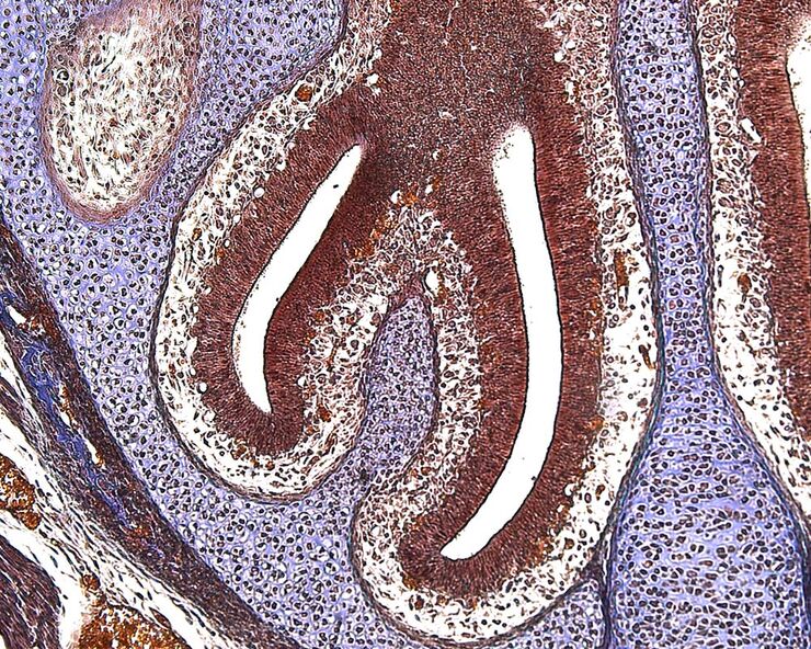
Brightfield image of a stained mouse embryo (brain)
Brightfield image of a stained mouse embryo (brain). Specimen courtesy of IGBMC, France.
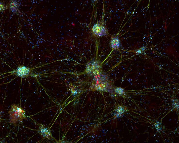
Cultured cortical neuronal cells (mouse). Simultaneous acquisition of 3 fluorochromes. Blue: DAP I, nuclei; Green: anti-Tubulin-Cy2; Red, Anti-Nestin-Cy3.
Image acquisition was performed using a Leica DFC7000 T/GT digital microscope camera in combination with a RGB cube in the microscope. Maximum projection done with LAS X software.
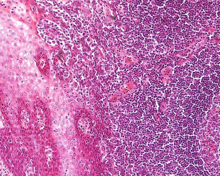
Brightfield image of H.E. stained human tissue
Brightfield image of H.E. stained human tissue. Acquired with Leica Application Suite X (LAS X)
