Optical filters and their spectral and physical characteristics
The Handbook continues with a thorough description of Chroma terminology and concepts used in the description of an optic and its spectral and physical characteristics. The difference between longpass/shortpass and bandpass filters is explained. The blocking/attenuation properties of filters are described and defined in optical density (OD). A brief overview is given regarding the importance of angle-of-incidence (AOI) and proper functioning of a given optic, followed by a consideration of cone-angle on these properties. The final portion of this section is a description of polarization effects on optics.
Traditional filter materials are illustrated, as well as more recent innovations to filter design and manufacture. Simple filters using colored glass, more advanced thin-film filter design and acousto-optical and liquid crystal tunable filters are all discussed, as are their relative advantages and disadvantages.
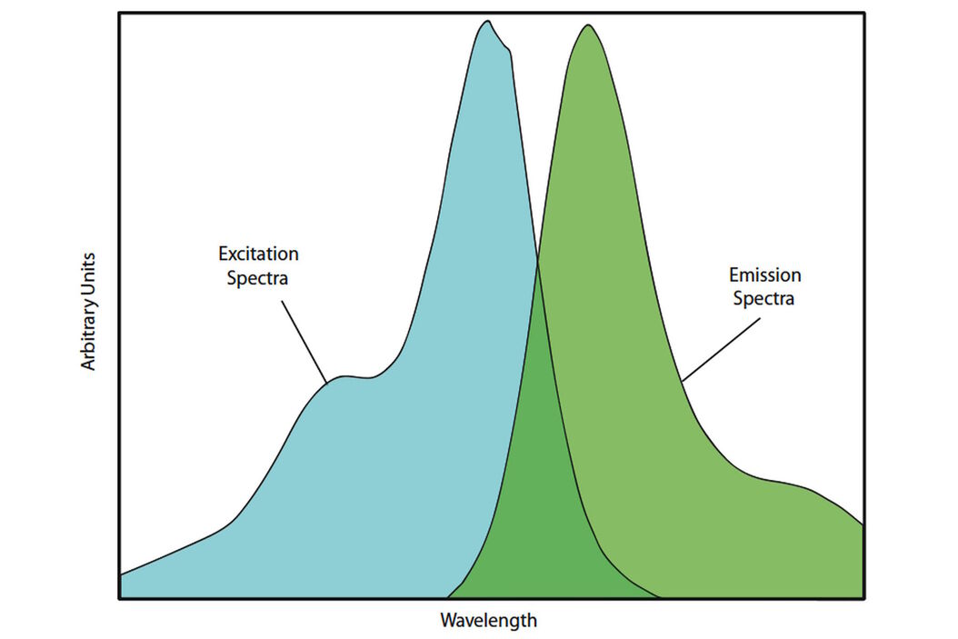
Fluorochromes, excitation and illumination sources
The next section contains a consideration of the factors that contribute to achieving excellent imaging results in a microscopy application. Choice of fluorochrome (either to be used alone or in concert with other fluorochromes), choice of excitation/illumination source and considerations of the quality of light reaching the detector are all discussed. Surface qualities (flatness, transmitted and reflected wavefront distortion and wedge, etc.) are reviewed and their effects on optimal beampath generation are described. Typical surface specifications are listed for Chroma-manufactured optics.
Image registration
Image-registration and visual overlap of multiple fluorescences within the microscope are considered. The ability to achieve sub-pixel registration of multiple images within the instrument is considered with a description of the effects of the various filters in this process.
Filters for confocal microscopes
A brief description of the filters used in confocal microscopy follows.
Also included is a glossary of terms that are used by Chroma to describe the optical properties of its filters, as well as numerous illustrations of the principles discussed.
Download
Download the complete “Handbook of Optical Filters for Fluorescence Microscopy”: http://www.chroma.com/sites/default/files/uploads/files/HandbookofOpticalFilters_0.pdf
Related Articles
-
")
Introduction to Fluorescent Proteins
Overview of fluorescent proteins (FPs) from, red (RFP) to green (GFP) and blue (BFP), with a table…
Sep 11, 2023Read article -

Diseases Linked to Scaffold Proteins and Signaling
This article shows how diseases related to scaffold proteins and protein signaling can be studied in…
Jan 23, 2023Read article -

Live-Cell Fluorescence Lifetime Multiplexing Using Organic Fluorophores
On-demand video: Imaging more subcellular targets by using fluorescence lifetime multiplexing…
Nov 18, 2022Read article
Related Pages
-
Fluorescence
Find out how fluorescence microscopes from Leica Microsystems support your research. Fluorescence is…
Visit related page
