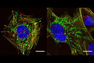
, actin network (ATTO 647N), and nuclear pore basket (CF 680R).")
, actin network (ATTO 647N), and nuclear pore basket (CF 680R).")
Related Articles
-

Extended Live-cell Imaging at Nanoscale Resolution
Extended live-cell imaging with TauSTED Xtend. Combined spatial and lifetime information allow…
Mar 05, 2024Read article -

Five-color FLIM-STED with One Depletion Laser
Webinar on five-color STED with a single depletion laser and fluorescence lifetime phasor…
Dec 14, 2022Read article -
A Versatile Palette of Fluorescent Probes
Researchers at the Max Planck Institute for Medical Research in Heidelberg have developed a general…
Jan 24, 2022Read article
Related Pages
-

Confocal Microscopes
Our confocal microscopes for top-class biomedical research provide imaging precision for subcellular…
Visit related page -
Super-Resolution
Find out more about Leica super-resolution microscopy solutions and how they can empower you to…
Visit related page
