
Science Lab
Science Lab
The knowledge portal of Leica Microsystems offers scientific research and teaching material on the subjects of microscopy. The content is designed to support beginners, experienced practitioners and scientists alike in their everyday work and experiments. Explore interactive tutorials and application notes, discover the basics of microscopy as well as high-end technologies – become part of the Science Lab community and share your expertise!
Filter articles
Tags
Story Type
Products
Loading...

Overcoming Observational Challenges in Organoid 3D Cell Culture
Learn how to overcome challenges in observing organoid growth. Read this article and discover new solutions for real-time monitoring which do not disturb the 3D structure of the organoids over time.
Loading...

Understanding Clearly the Magnification of Microscopy
To help users better understand the magnification of microscopy and how to determine the useful range of magnification values for digital microscopes, this article provides helpful guidelines.
Loading...
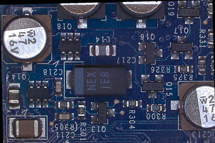
Rapid and Reliable Examination of PCBs & PCBAs with Digital Microscopy
Digital microscopes provide users with a convenient and rapid way to acquire high-quality, reliable image data and make quick inspection and analysis of printed circuit boards (PCBs) and assemblies…
Loading...

Digital Inspection Microscope for Industrial Applications
Factors users should consider before choosing a digital inspection microscope for industrial applications, including quality control (QC), failure analysis (FA), and R&D, are described in this…
Loading...
 taken with a ring light (RL) and near vertical illumination (NVI).")
Microscope Illumination for Industrial Applications
Inspection microscope users can obtain information from this article which helps them choose the optimal microscope illumination or lighting system for inspection of parts or components.
Loading...

How to Benefit from Digital Cytopathology
If you have thought of digital cytopathology as characterized by the digitization of glass slides, this webinar with Dr. Alessandro Caputo from the University Hospital of Salerno, Italy will broaden…
Loading...
Tracking Single Cells Using Deep Learning
AI-based solutions continue to gain ground in the field of microscopy. From automated object classification to virtual staining, machine and deep learning technologies are powering scientific…
Loading...

Learning the Cellular Architecture from its Optical Properties
In the last 3 years, microscopists have started to use "AI based" solutions for a wide range of applications, including image acquisition optimization (smart microscopy), object classification, image…
Loading...
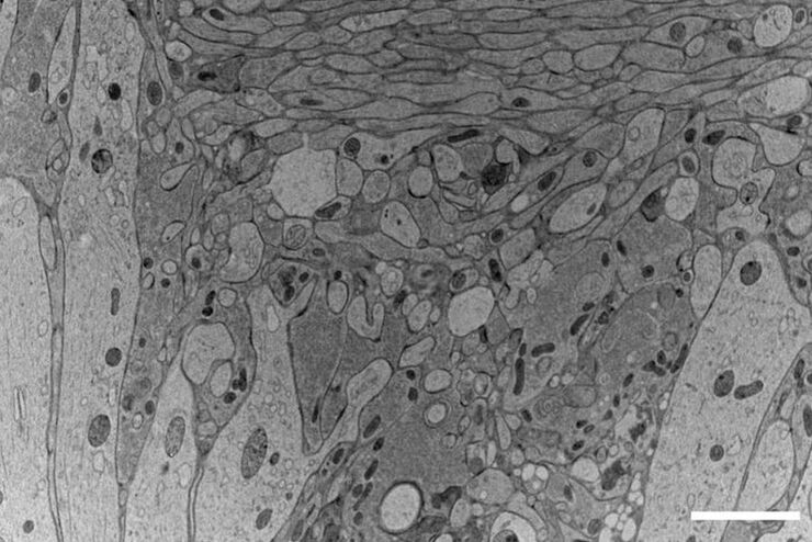
Workflows and Instrumentation for Cryo-electron Microscopy
Cryo-electron microscopy is an increasingly popular modality to study the structures of macromolecular complexes and has enabled numerous new insights in cell biology. In recent years, cryo-electron…