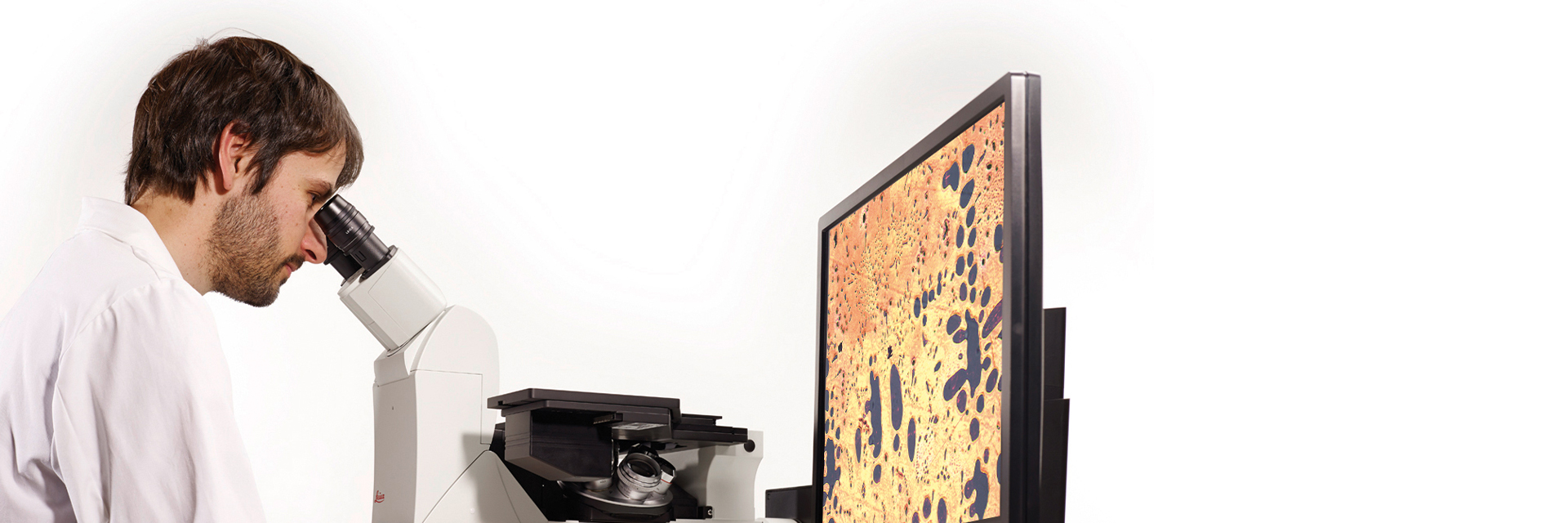
DIC Microscopes
Most people use brightfield illumination when using a microscope. Unfortunately, it usually provides just a low-contrast image of many biological specimens where few details can be distinguished. Selective stains can be used to enhance the contrast with brightfield, but there is the risk that the stains can be toxic to living cells. With DIC microscopy, where DIC means differential interference contrast, no stains are needed to see the structures of many types of biological specimens with greater contrast compared to brightfield. The contrast method exploits polarized light and differences in thickness or optical density of the specimen that lead to a phase shift of the light passing through it.
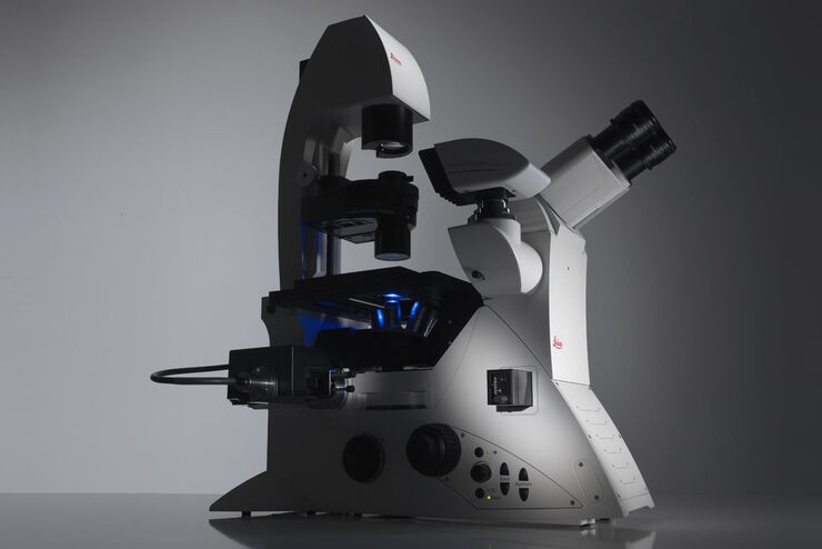
DIC is offered with specific Leica microscopes. It can be useful for the observation of cells or tissues in a variety of life-science or forensic applications. DIC microscopy can also be helpful for certain material and mineral applications.
Contact us for expert advice on the right dic microscope for your needs and budget.

What is DIC microscopy and what is it used for?
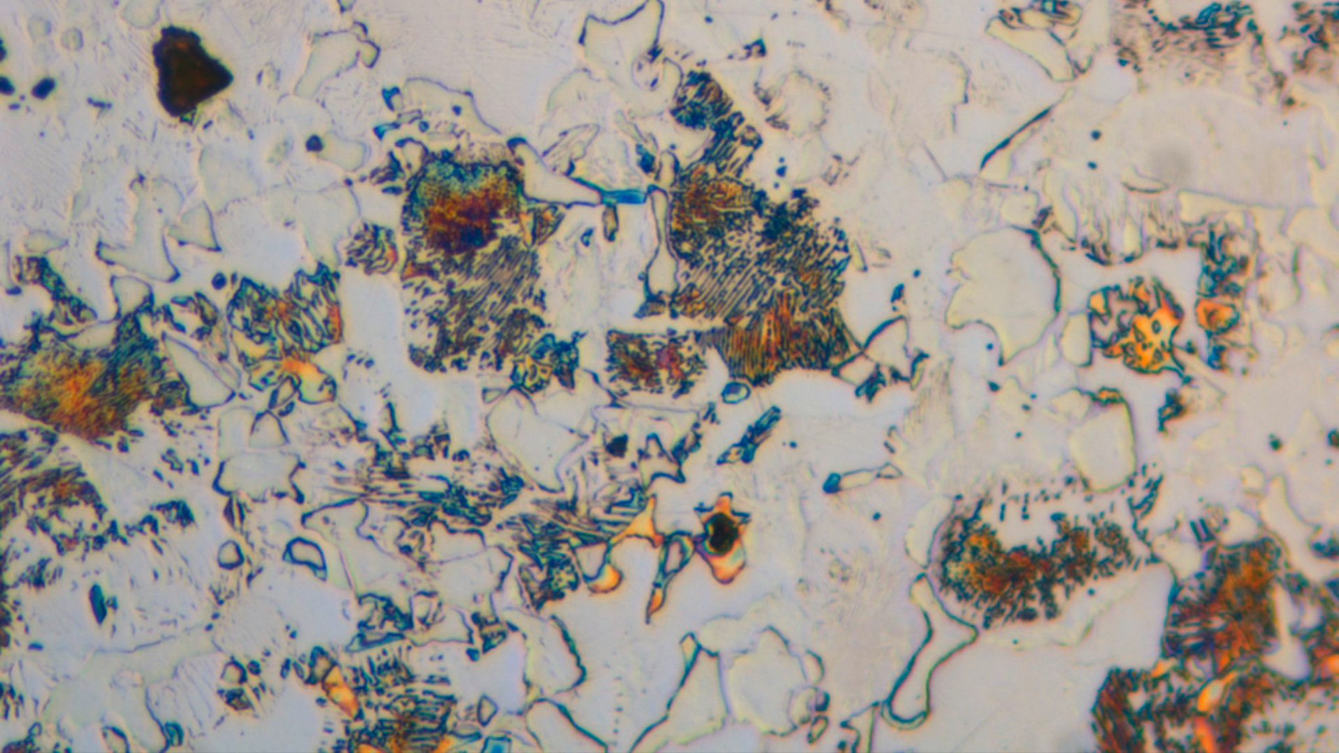
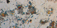
DIC is differential interference contrast, an optical contrast technique for microscopy which allows unstained structures in the cells of biological specimens to be observed with adequate contrast and resolution. It can also help to visualize small height differences on the surfaces of materials. Only polarized light is used to illuminate the specimen. The polarized light is dispersed into two distinct light rays with an orthogonal plane of polarization. As the rays experience different refraction or scattering in the specimen, different phase shifts occur. When these light rays reunite, they interfere and become elliptically polarized. This polarization can be changed into an amplitude shift via an analyzer. Phase shifts corresponding to differences as small as 1/1000 of a wavelength (when detected with a camera sensor) can be observed in the DIC images. DIC microscopy images are relief-like and seem to have a shadow.

How do you set up DIC microscopy?
A DIC microscope is a widefield microscopy which has a polarization filter and Wollaston prism between the light source and condenser lens and also between the objective lens and camera sensor or eyepieces. For more information about how it is set up, refer to the articles: Differential Interference Contrast (DIC) & Metallography

How does differential interference contrast (DIC) microscopy work?
A DIC microscope is a conventional widefield microscope which uses polarization filters and Wollaston prisms.
Light from a source passes through a filter and becomes 45° polarized. After passing through a Wollaston prism, the light is separated into perpendicular polarized components where one is 0° and the other 90° polarized. The condenser lens focusses the light onto the specimen which is illuminated by 2 coherent parallel rays with different polarization. The rays experience different optical path lengths, as there may be differences in thickness or refractive index along the path where they pass through the specimen.
The result is a phase shift of one ray compared to the other. After passing through the objective and another Wollaston prism, the perpendicularly polarized light is recombined to become one which is 135° polarized. Depending on the differences in the optical path lengths, interference of the 2 rays results in constructive (brightening) or destructive (darkening) interference, making what were hardly visible structures with brightfield now observable with DIC.
Finally, directly transmitted light, which did not experience any phase shift, is removed by the polarization filter (also called analyzer) which only lets 135°-polarized light pass. For more details, refer to the article: Differential Interference Contrast (DIC)
Frequently Asked Questions DIC Microscopes
A DIC microscope is similar to a conventional brightfield microscope, except it uses a polarization filter and Wollaston prism between the light source and condenser lens and also between the objective and camera sensor or eyepieces. For more information, refer to the articles: Differential Interference Contrast (DIC) & Metallography
In the early 1950s, Georges Nomarski invented the microscopy contrast technique called differential interference contrast or DIC. Today, it is still a widely used technique. For more details, refer to the articles: A Brief History of Light Microscopy & Differential Interference Contrast (DIC)
Yes, a DIC microscope can be equipped with a camera for recording DIC images observed with the contrast method.
Related Articles
 and oblique (right) brightfield illumination using a Leica compound microscope. The defect on the wafer surface is clearly more visible with oblique illumination.")
Rapid Semiconductor Inspection with Microscope Contrast Methods

Differential Interference Contrast (DIC) Microscopy
Factors to Consider When Selecting a Research Microscope

Introduction to Mammalian Cell Culture

Introduction to Widefield Microscopy
 inoculated with cowpea mosaic virus (CPMV) containing the GFP-gene inserted between the movement protein (MP) and the capsid proteins (CPs) in the viral RNA 2")
Introduction to Live-Cell Imaging

Optical Contrast Methods

Interested to know more?
Talk to our experts.
Do you prefer personal consulting? Show local contacts