
Science Lab
Science Lab
Das Wissensportal von Leica Microsystems bietet Ihnen Wissens- und Lehrmaterial zu den Themen der Mikroskopie. Die Inhalte sind so konzipiert, dass sie Einsteiger, erfahrene Praktiker und Wissenschaftler gleichermaßen bei ihrem alltäglichen Vorgehen und Experimenten unterstützen. Entdecken Sie interaktive Tutorials und Anwendungsberichte, erfahren Sie mehr über die Grundlagen der Mikroskopie und High-End-Technologien - werden Sie Teil der Science Lab Community und teilen Sie Ihr Wissen!
Loading...

Technical Terms for Digital Microscope Cameras and Image Analysis
Learn more about the basic principles behind digital microscope camera technologies, how digital cameras work, and take advantage of a reference list of technical terms from this article.
Loading...

Understanding Clearly the Magnification of Microscopy
To help users better understand the magnification of microscopy and how to determine the useful range of magnification values for digital microscopes, this article provides helpful guidelines.
Loading...

ISO 9022 Standard Part 11 - Testing Microscopes with Severe Conditions
This article describes a test to determine the robustness of Leica microscopes to mold and fungus growth. The test follows the specifications of the ISO 9022 part 11 standard for optical instruments.
Loading...

Life-Science-Forschung: Welche Mikroskopkamera ist die richtige für Sie?
Wie Sie entscheiden, welche Kamera für Ihre Life-Science-Mikroskopie-Experimente die Richtige ist. Welche Kamera von Leica Microsystems ist für Sie am besten geeignet?
Loading...

Differential Interference Contrast (DIC) Microscopy
This article demonstrates how differential interference contrast (DIC) can be actually better than brightfield illumination when using microscopy to image unstained biological specimens.
Loading...
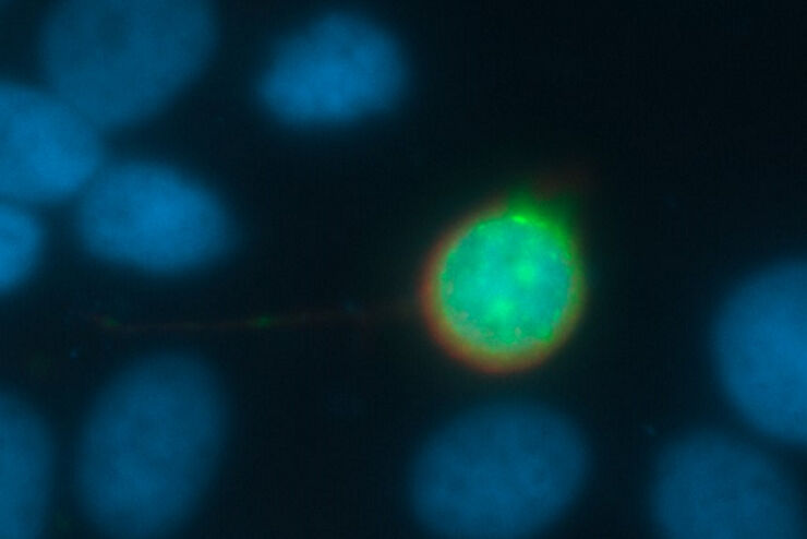
Going Beyond Deconvolution
Widefield fluorescence microscopy is often used to visualize structures in life science specimens and obtain useful information. With the use of fluorescent proteins or dyes, discrete specimen…
Loading...
 cells taken with phase contrast.")
Phase Contrast and Microscopy
This article explains phase contrast, an optical microscopy technique, which reveals fine details of unstained, transparent specimens that are difficult to see with common brightfield illumination.
Loading...
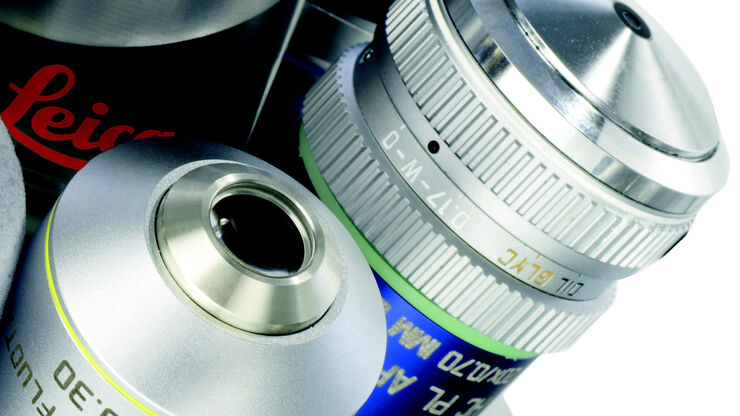
Immersion Objectives
How an immersion objective, which has a liquid medium between it and the specimen being observed, helps increase the numerical aperture and microscope resolution is explained in this article.
Loading...
 of an image of two points where the distance between them corresponds to the Rayleigh criterion.")
Microscope Resolution: Concepts, Factors and Calculation
This article explains in simple terms microscope resolution concepts, like the Airy disc, Abbe diffraction limit, Rayleigh criterion, and full width half max (FWHM). It also discusses the history.