
Science Lab
Science Lab
Das Wissensportal von Leica Microsystems bietet Ihnen Wissens- und Lehrmaterial zu den Themen der Mikroskopie. Die Inhalte sind so konzipiert, dass sie Einsteiger, erfahrene Praktiker und Wissenschaftler gleichermaßen bei ihrem alltäglichen Vorgehen und Experimenten unterstützen. Entdecken Sie interaktive Tutorials und Anwendungsberichte, erfahren Sie mehr über die Grundlagen der Mikroskopie und High-End-Technologien - werden Sie Teil der Science Lab Community und teilen Sie Ihr Wissen!
Loading...

Overcoming Observational Challenges in Organoid 3D Cell Culture
Learn how to overcome challenges in observing organoid growth. Read this article and discover new solutions for real-time monitoring which do not disturb the 3D structure of the organoids over time.
Loading...

How to Streamline Your Histology Workflows
Streamline your histology workflows. The unique Fluosync detection method embedded into Mica enables high-res RGB color imaging in one shot.
Loading...
Accelerating Discovery for Multiplexed Imaging of Diverse Tissues
Explore IBEX: Open-source multiplexed imaging. Join the collaborative IBEX Imaging Community for optimized tissue processing, antibody selection, and human atlas construction.
Loading...

Notable AI-based Solutions for Phenotypic Drug Screening
Learn about notable optical microscope solutions for phenotypic drug screening using 3D-cell culture, both planning and execution, from this free, on-demand webinar.
Loading...

Understanding Tumor Heterogeneity with Protein Marker Imaging
Explore tumor heterogeneity and immune cell dynamics. See how quantitative imaging analysis reveals spatial relationships and molecular insights crucial for advancing cancer research and therapeutics.
Loading...
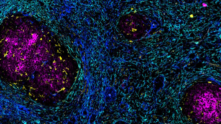
![[Translate to German:] Co-detection of 10 extracellular matrix proteins and 3 topographical tissue landmarks by multiplex immunostaining within a single high-grade fibrous hotspot from a human hepatocellular carcinoma](/fileadmin/_processed_/0/8/csm_Single_high-grade_fibrous_hotspot_from_a_human_hepatocellular_carcinoma_e5541282bd.jpg "[Translate to German:] Co-detection of 10 extracellular matrix proteins and 3 topographical tissue landmarks by multiplex immunostaining within a single high-grade fibrous hotspot from a human hepatocellular carcinoma")
In-situ-Identifizierung von Krebsstammzellnischen im Leberzellkarzinom
Die Multiplex-Bildgebung spielt eine entscheidende Rolle bei der Krebsforschung, da sie es Wissenschaftlern ermöglicht, tief in die komplexe Mikroumgebung von Tumoren einzudringen und einzigartige…
Loading...

Multiplexing with Luke Gammon: Advance your Spatial Biology Research
Learn how multiplexing imaging and spatial biology can help researchers better understand complex biological systems. In this interview, Dr. Gammon and Dr. Pointu of Leica Microsystems discuss pain…
Loading...

How to Determine Cell Confluency with a Digital Microscope
This article shows how to measure cell confluency in an easy and consistent way with Mateo TL, increasing confidence in downstream experiments.
Loading...

How to do a Proper Cell Culture Quick Check
In order to successfully work with mammalian cell lines, they must be grown under controlled conditions and require their own specific growth medium. In addition, to guarantee consistency their growth…