
Science Lab
Science Lab
Das Wissensportal von Leica Microsystems bietet Ihnen Wissens- und Lehrmaterial zu den Themen der Mikroskopie. Die Inhalte sind so konzipiert, dass sie Einsteiger, erfahrene Praktiker und Wissenschaftler gleichermaßen bei ihrem alltäglichen Vorgehen und Experimenten unterstützen. Entdecken Sie interaktive Tutorials und Anwendungsberichte, erfahren Sie mehr über die Grundlagen der Mikroskopie und High-End-Technologien - werden Sie Teil der Science Lab Community und teilen Sie Ihr Wissen!
Loading...
")
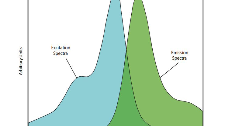
Introduction to Fluorescent Proteins
Overview of fluorescent proteins (FPs) from, red (RFP) to green (GFP) and blue (BFP), with a table showing their relevant spectral characteristics.
Loading...

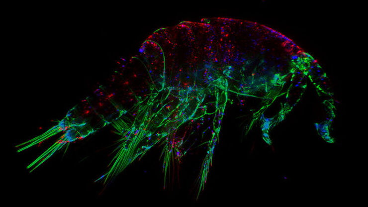
Diseases Linked to Scaffold Proteins and Signaling
This article shows how diseases related to scaffold proteins and protein signaling can be studied in zebrafish models efficiently with a THUNDER Imager.
Loading...

Live-Cell Fluorescence Lifetime Multiplexing Using Organic Fluorophores
On-demand video: Imaging more subcellular targets by using fluorescence lifetime multiplexing combined with spectrally resolved detection.
Loading...
 and THUNDER image (bottom).")
The Neural Crest (NC)
This article discusses how the study of neural crest (NC) development in chicken embryos is aided with haze-free imaging using a THUNDER Imager 3D Assay. Proper specification, migration, and…
Loading...
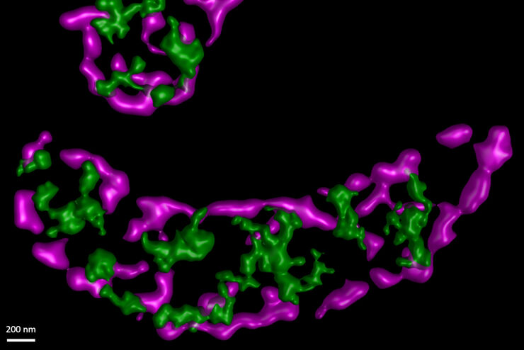
STED Sheds Insight on Mitochondrial Protein Synthesis
In this paper the authors have adapted a click chemistry-based method coupled with stimulated emission depletion nanoscopy (STED) to address these questions reporting that, in human cells in culture,…
Loading...
The Fundamentals and History of Fluorescence and Quantum Dots
At some point in your research and science career, you will no doubt come across fluorescence microscopy. This ubiquitous technique has transformed the way in which microscopists can image, tag and…
Loading...

Photoactivatable, Photoconvertible, and Photoswitchable Fluorescent Proteins
Fluorescent proteins (FPs) such as GFP, YFP or DsRed are powerful tools to visualize cellular components in living cells. Nevertheless, there are circumstances when classical FPs reach their limits.…
Loading...
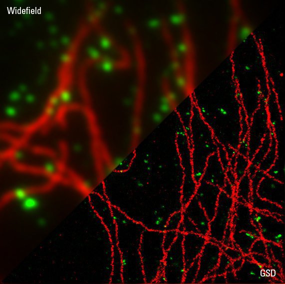
Sample Preparation for GSDIM Localization Microscopy – Protocols and Tips
The widefield super-resolution technique GSDIM (Ground State Depletion followed by individual molecule return) is a localization microscopy technique that is capable of resolving details as small as…
Loading...
Handbook of Optical Filters for Fluorescence Microscopy
Fluorescence microscopy and other light-based applications require optical filters that have demanding spectral and physical characteristics. Often, these characteristics are application-specific and…