
Science Lab
Science Lab
Das Wissensportal von Leica Microsystems bietet Ihnen Wissens- und Lehrmaterial zu den Themen der Mikroskopie. Die Inhalte sind so konzipiert, dass sie Einsteiger, erfahrene Praktiker und Wissenschaftler gleichermaßen bei ihrem alltäglichen Vorgehen und Experimenten unterstützen. Entdecken Sie interaktive Tutorials und Anwendungsberichte, erfahren Sie mehr über die Grundlagen der Mikroskopie und High-End-Technologien - werden Sie Teil der Science Lab Community und teilen Sie Ihr Wissen!
Loading...

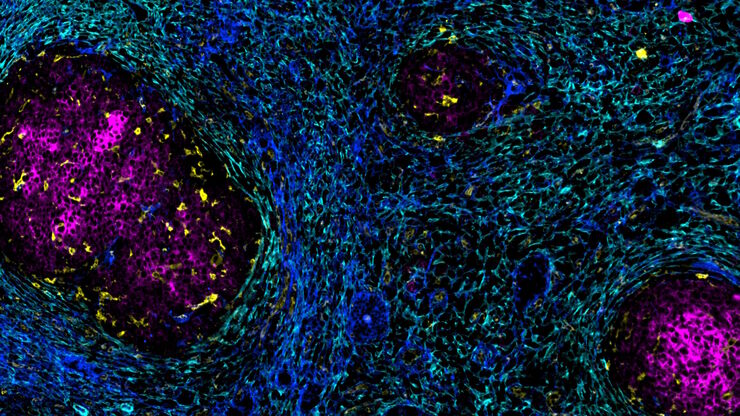
IBEX, Cell DIVE, and RNA-Seq: A Multi-omics Approach to Follicular Lymphoma
In a recent study by Radtke et al., a multi-omics spatial biology approach helps shed light on early relapsing lymphoma patients
Loading...
Accelerating Discovery for Multiplexed Imaging of Diverse Tissues
Explore IBEX: Open-source multiplexed imaging. Join the collaborative IBEX Imaging Community for optimized tissue processing, antibody selection, and human atlas construction.
Loading...

Transforming Multiplexed 2D Data into Spatial Insights Guided by AI
Aivia 13 handles large 2D images and enables researchers to obtain deep insights into microenvironment surrounding their phenotypes with millions of detected objects and automatic clustering up to 30…
Loading...

Understanding Tumor Heterogeneity with Protein Marker Imaging
Explore tumor heterogeneity and immune cell dynamics. See how quantitative imaging analysis reveals spatial relationships and molecular insights crucial for advancing cancer research and therapeutics.
Loading...

The Shape of the Brain: Spatial Biology of Alzheimer’s Disease
Uncover cell identity and brain structure in Alzheimer's disease with Cell DIVE multiplexed imaging, demonstrating how spatial biology can lead to advances in therapy development for…
Loading...
![[Translate to German:] Co-detection of 10 extracellular matrix proteins and 3 topographical tissue landmarks by multiplex immunostaining within a single high-grade fibrous hotspot from a human hepatocellular carcinoma](/fileadmin/_processed_/0/8/csm_Single_high-grade_fibrous_hotspot_from_a_human_hepatocellular_carcinoma_e5541282bd.jpg "[Translate to German:] Co-detection of 10 extracellular matrix proteins and 3 topographical tissue landmarks by multiplex immunostaining within a single high-grade fibrous hotspot from a human hepatocellular carcinoma")
In-situ-Identifizierung von Krebsstammzellnischen im Leberzellkarzinom
Die Multiplex-Bildgebung spielt eine entscheidende Rolle bei der Krebsforschung, da sie es Wissenschaftlern ermöglicht, tief in die komplexe Mikroumgebung von Tumoren einzudringen und einzigartige…
Loading...

Discover how Multiplexed Bioimaging can Advance Cancer Research
Explore multiplexing with up to 60 biomarkers, enabling advanced tumor imaging approaches to gather precise, spatially-resolved single-cell data that helps enhance cancer research and clinical…
Loading...

Potential of Multiplex Confocal Imaging for Cancer Research and Immunology
Explore the new frontiers of multi-color fluorescent imaging: from image acquisition to analysis
Loading...

Multiplexing with Luke Gammon: Advance your Spatial Biology Research
Learn how multiplexing imaging and spatial biology can help researchers better understand complex biological systems. In this interview, Dr. Gammon and Dr. Pointu of Leica Microsystems discuss pain…