
Science Lab
Science Lab
Das Wissensportal von Leica Microsystems bietet Ihnen Wissens- und Lehrmaterial zu den Themen der Mikroskopie. Die Inhalte sind so konzipiert, dass sie Einsteiger, erfahrene Praktiker und Wissenschaftler gleichermaßen bei ihrem alltäglichen Vorgehen und Experimenten unterstützen. Entdecken Sie interaktive Tutorials und Anwendungsberichte, erfahren Sie mehr über die Grundlagen der Mikroskopie und High-End-Technologien - werden Sie Teil der Science Lab Community und teilen Sie Ihr Wissen!
Loading...

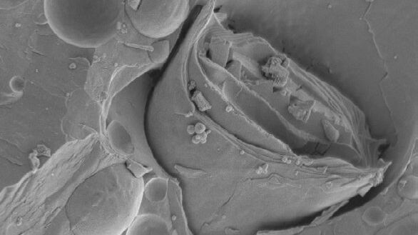
Wie die Analyse von Meeresmikroorganismen durch Hochdruckgefrieren verbessert werden kann
Die ultrastrukturelle Analyse von Umweltproben, hier Dinoflagellaten, bleibt heutzutage eine Herausforderung. Hier zeigen wir, dass die Durchführung von Hochdruckgefrieren (HPF) vor Ort die…
Loading...
Advancing Cellular Ultrastructure Research
Freeze-fracture and freeze-etching are useful tools for studying flexible membrane-associated structures such as tight junctions or the enteric glycocalyx. Freeze-fracture and etching are two…
Loading...

Targeting Active Recycling Nuclear Pore Complexes using Cryo Confocal Microscopy
In this article, how cryo light microscopy and, in particular cryo confocal microscopy, is used to improve the reliability of cryo EM workflows is described. The quality of the EM grids and samples is…
Loading...

Introduction to Ultramicrotomy
When studying samples, to visualize their fine structure with nanometer scale resolution, most often electron microscopy is used. There are 2 types: scanning electron microscopy (SEM) which images the…
Loading...

High Resolution Array Tomography with Automated Serial Sectioning
The optimization of high resolution, 3-dimensional (3D), sub-cellular structure analysis with array tomography using an automated serial sectioning solution, achieving a high section density on the…
Loading...
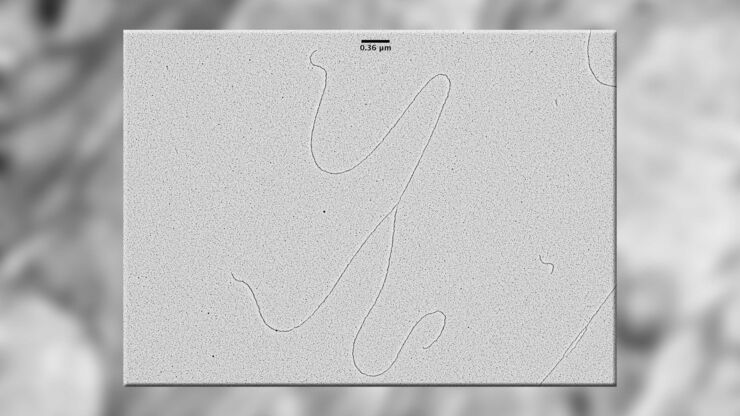
Visualization of DNA Molecules
Precise low angle rotary shadowing with heavy metals (like platinum) can be used in transmission electron microscopy (TEM) to observe molecular details of objects previously absorbed on a thin, low…
Loading...
Ion Beam Polishing of Sample Surfaces - Sample Preparation for SEM
Application Note for Leica EM RES102 - Ion milling can be used to reduce the roughness of sample surfaces. Small angles less than 6° with respect to the sample surface are necessary. The high voltage…
Loading...
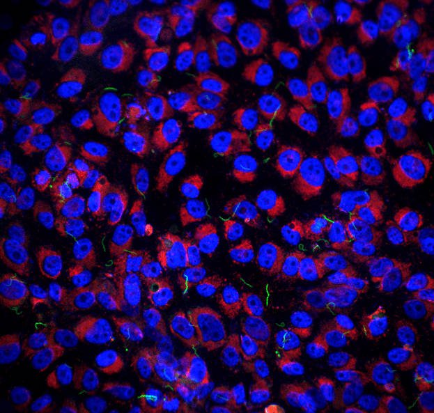
Imaging of Host Cell-bacteria Interactions using Correlative Microscopy under Cryo-conditions
Pathogenic bacteria have developed intriguing strategies to establish and promote infections in their respective hosts. Most bacterial pathogens initiate infectious diseases by adhering to host cells…
Loading...
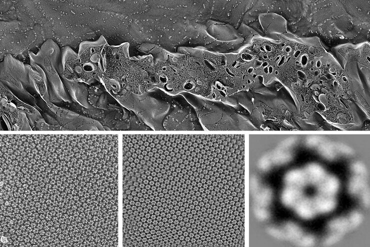
Brief Introduction to Freeze Fracture and Etching
Freeze fracture describes the technique of breaking a frozen specimen to reveal internal structures. Freeze etching is the sublimation of surface ice under vacuum to reveal details of the fractured…