THUNDER Imager Tissue
THUNDER Imaging Systems
Prodotti
Home
Leica Microsystems
THUNDER Imager Tissue
Decodifica la biologia 3D in tempo reale*
Leggi gli articoli più recenti
 acquired using THUNDER Imager Live Cell. Image courtesy of Janina Kaspar and Irene Santisteban, Schäfer Lab, TUM.")
Imaging Organoid Models to Investigate Brain Health
Imaging human brain organoid models to study the phenotypes of specialized brain cells called microglia, and the potential applications of these organoid models in health and disease.
. Image courtesy of Prof. Hui Guo, School of Life Sciences, Central South University, China")
How Microscopy Helps the Study of Mechanoceptive and Synaptic Pathways
In this podcast, Dr Langenhan explains how microscopy helps his team to study mechanoceptive and synaptic pathways, their challenges, and how they overcome them.

What are the Challenges in Neuroscience Microscopy?
eBook outlining the visualization of the nervous system using different types of microscopy techniques and methods to address questions in neuroscience.
Going Beyond Deconvolution
Widefield fluorescence microscopy is often used to visualize structures in life science specimens and obtain useful information. With the use of fluorescent proteins or dyes, discrete specimen…

Fast, High Acuity Imaging and AI-assisted Analysis
The use of state-of-the-art AI systems is pushing image analysis into a new generation. Challenges like the conflict between imaging power and sample integrity are being overcome with THUNDER’s…

Create New Options for Live Cell Imaging
The use of state-of-the-art AI systems is pushing image analysis into a new generation. Challenges like the conflict between imaging power and sample integrity are being overcome with THUNDER’s…
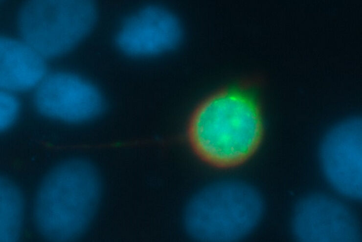
 expressed in the sensory neurons.")
Fast, High-contrast 3D Imaging of Sensory Neurons
This article discusses how fast, high-contrast 3D imaging of dorsal root ganglion (DRG) tissue with a THUNDER Imager Tissue using large volume computational clearing (LVCC) allows sensory neurons to…

Accurately Analyze Fluorescent Widefield Images
The specificity of fluorescence microscopy allows researchers to accurately observe and analyze biological processes and structures quickly and easily, even when using thick or large samples. However,…
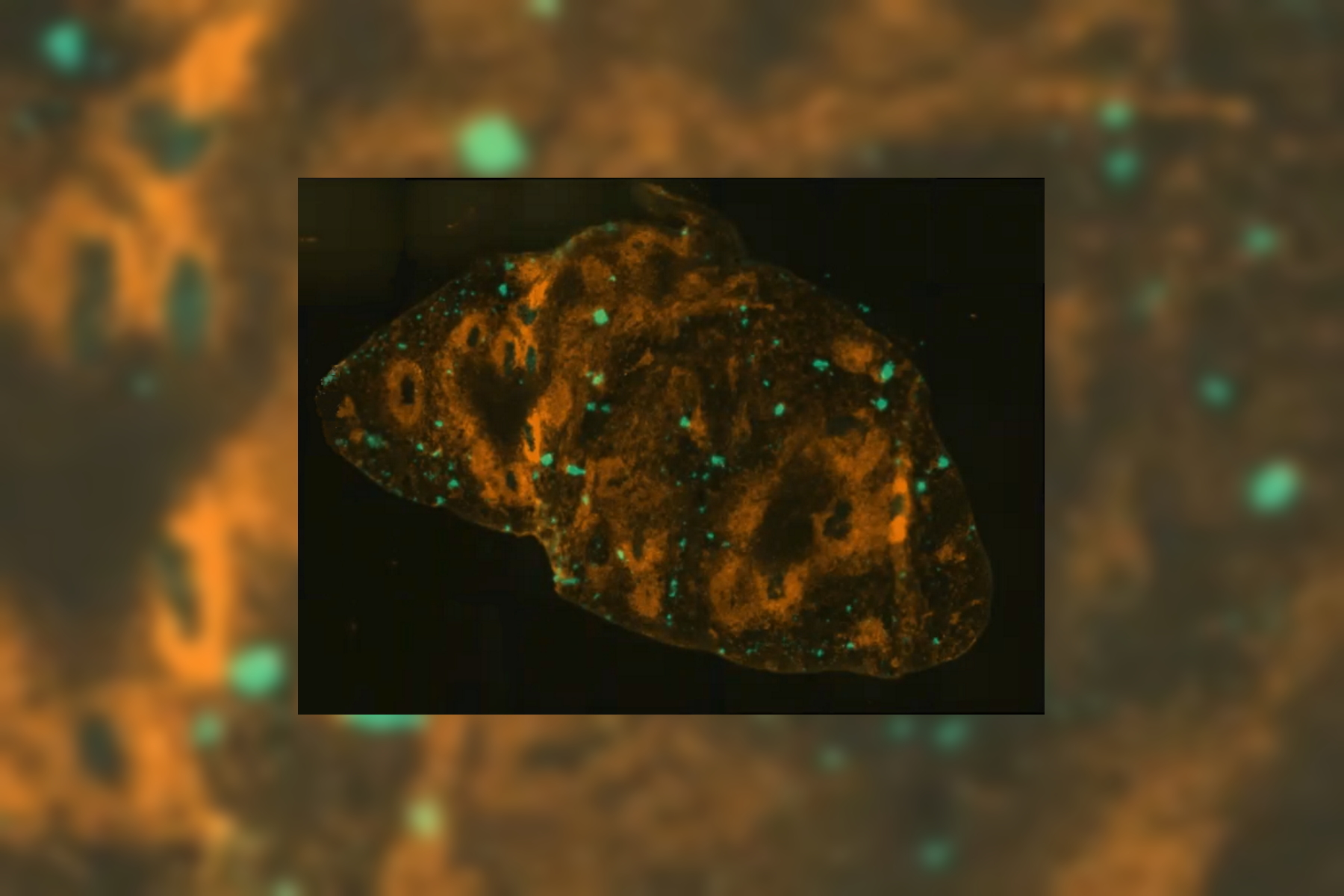
 and THUNDER (right) image of Ewing Sarcoma cells (SK-ES-1)")
Visualizing the Mitotic Spindle in Cancer Cells
This article demonstrates how this research is aided by visualizing more details of mitotic spindles in Ewing Sarcoma cells using the THUNDER Imager Tissue and Large Volume Computational Clearing…

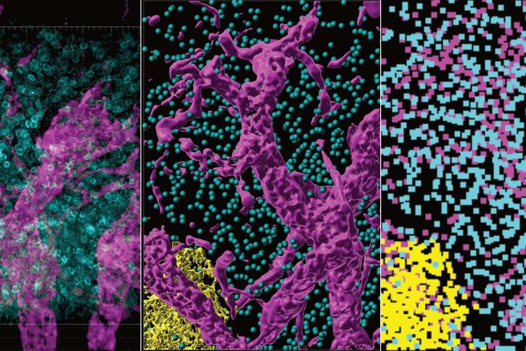
High-resolution 3D Imaging to Investigate Tissue Ageing
Award-winning researcher Dr. Anjali Kusumbe demonstrates age-related changes in vascular microenvironments through single-cell resolution 3D imaging of young and aged organs.
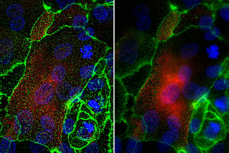
Role of Mucins and Glycosylation in Dry Eye Disease
This article shows how fast, high-contrast, and sharp imaging of stratified human corneal epithelial cells with THUNDER imaging technology for dry eye disease (DED) research allows membrane ridges to…
Optimizing THUNDER Platform for High-Content Slide Scanning
With rising demand for full-tissue imaging and the need for FL signal quantitation in diverse biological specimens, the limits on HC imaging technology are tested, while user trainability and…

Physiology Image Gallery
Physiology is about the processes and functions within a living organism. Research in physiology focuses on the activities and functions of an organism’s organs, tissues, or cells, including the…

Developmental Biology Image Gallery
Developmental biology explores the development of complex organisms from the embryo to adulthood to understand in detail the origins of disease. This category of the gallery shows images about…
, specific neuronal markers (magenta) and cell nuclei (white), computational cleared.")
Into the Third Dimension with "Wow Effect"- Observe Cells in 3D and Real-Time
Life is fast, especially for a cell. As a rule, cells should be examined under physiological conditions which are as close as possible to their natural environment. New technologies offer tremendous…

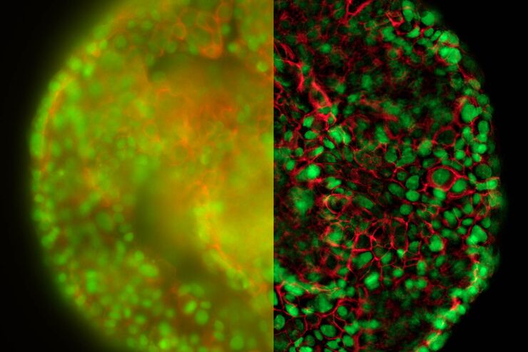
Observing 3D Cell Cultures During Development
3D cell cultures, such as organoids and spheroids, give insights into cells and their interactions with their microenvironment. These 3D cell cultures are playing an increasingly important role for…
 and cytokinesis ring (Rlc1-mCherry; red).")
Studying Cell Division
Cell division is a biological process during which all cellular components must be distributed among the daughter cells. The division process requires firm coordination for success. Microscopy is…

The Power of Pairing Adaptive Deconvolution with Computational Clearing
Learn how deconvolution allows you to overcome losses in image resolution and contrast in widefield fluorescence microscopy due to the wave nature of light and the diffraction of light by optical…
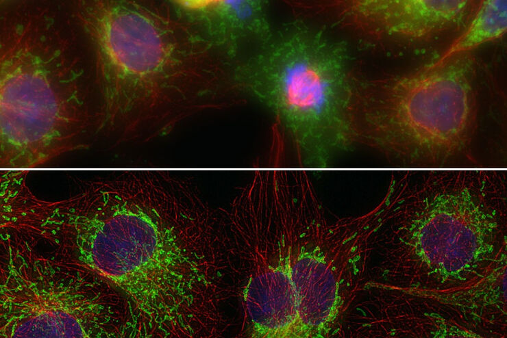
Improvement of Imaging Techniques to Understand Organelle Membrane Cell Dynamics
Understanding cell functions in normal and tumorous tissue is a key factor in advancing research of potential treatment strategies and understanding why some treatments might fail. Single-cell…

Image Gallery: THUNDER Imager
To help you answer important scientific questions, THUNDER Imagers eliminate the out-of-focus blur that clouds the view of thick samples when using camera-based fluorescence microscopes. They achieve…
From Organs to Tissues to Cells: Analyzing 3D Specimens with Widefield Microscopy
Obtaining high-quality data and images from thick 3D samples is challenging using traditional widefield microscopy because of the contribution of out-of-focus light. In this webinar, Falco Krüger…

Studying Human Brain Development and Disease
Neural spheroids created from human induced pluripotent stem cells (iPSCs) provide effective and novel tools for studying brain development, as well as the underlying pathological mechanisms of…

An Introduction to Computational Clearing
Many software packages include background subtraction algorithms to enhance the contrast of features in the image by reducing background noise. The most common methods used to remove background noise…
Factors to Consider When Selecting a Research Microscope
An optical microscope is often one of the central devices in a life-science research lab. It can be used for various applications which shed light on many scientific questions. Thereby the…
Computational Clearing - Enhance 3D Specimen Imaging
This webinar is designed to clarify crucial specifications that contribute to THUNDER Imagers' transformative visualization of 3D samples and improvements within a researcher's imaging-related…
THUNDER Imagers: High Performance, Versatility and Ease-of-Use for your Everyday Imaging Workflows
This webinar will showcase the versatility and performance of THUNDER Imagers in many different life science applications: from counting nuclei in retina sections and RNA molecules in cancer tissue…
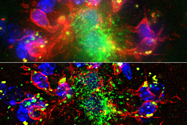
Evaluating Axon Regeneration After Brain or Spine Trauma of Mice
Damaged nerve regeneration was investigated using mouse spinal cord sections treated with compounds that counter axon growth inhibitor (AGI) proteins. The sections were screened to find active and…
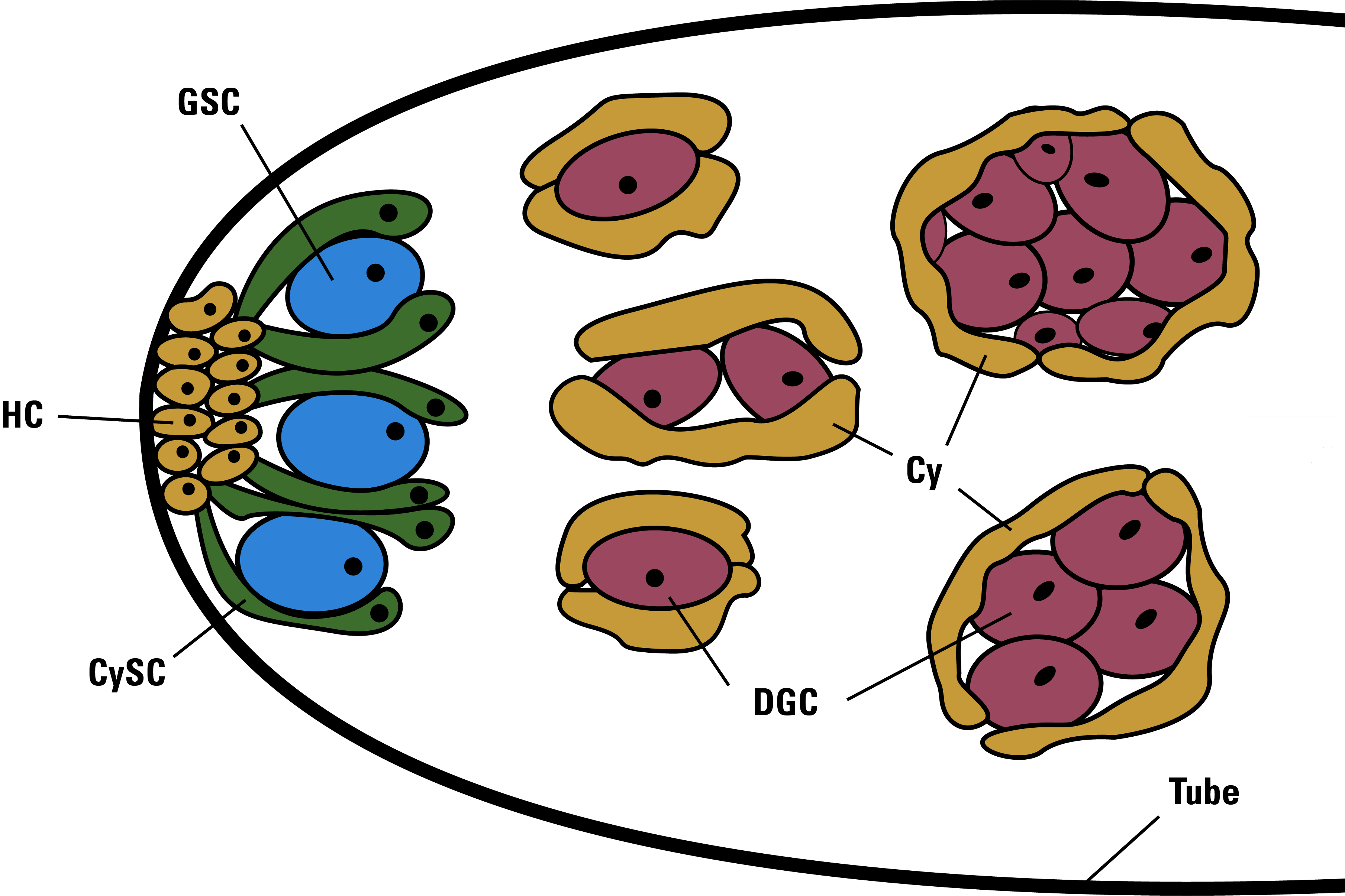
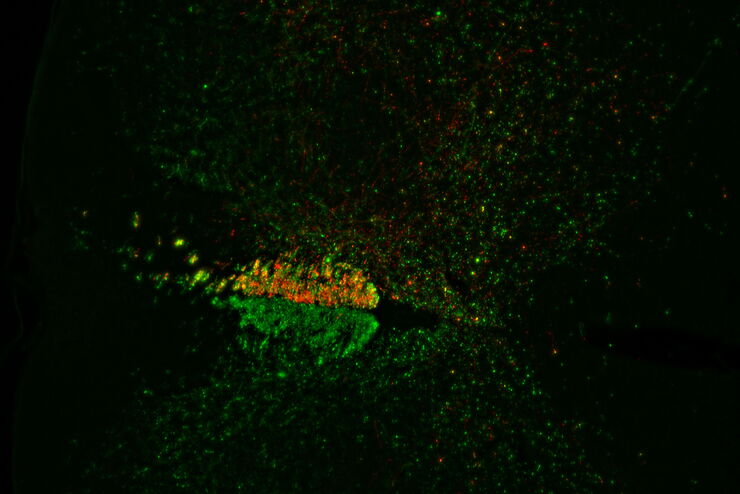
Drosophila Testis Niche Stem Cells – Three Color Computational Clearing
Differentiated living beings such as humans, but also a fruit fly or a plant, possess not only the differentiated cells which form specific tissues, but also those cells whose fate is not yet (or only…
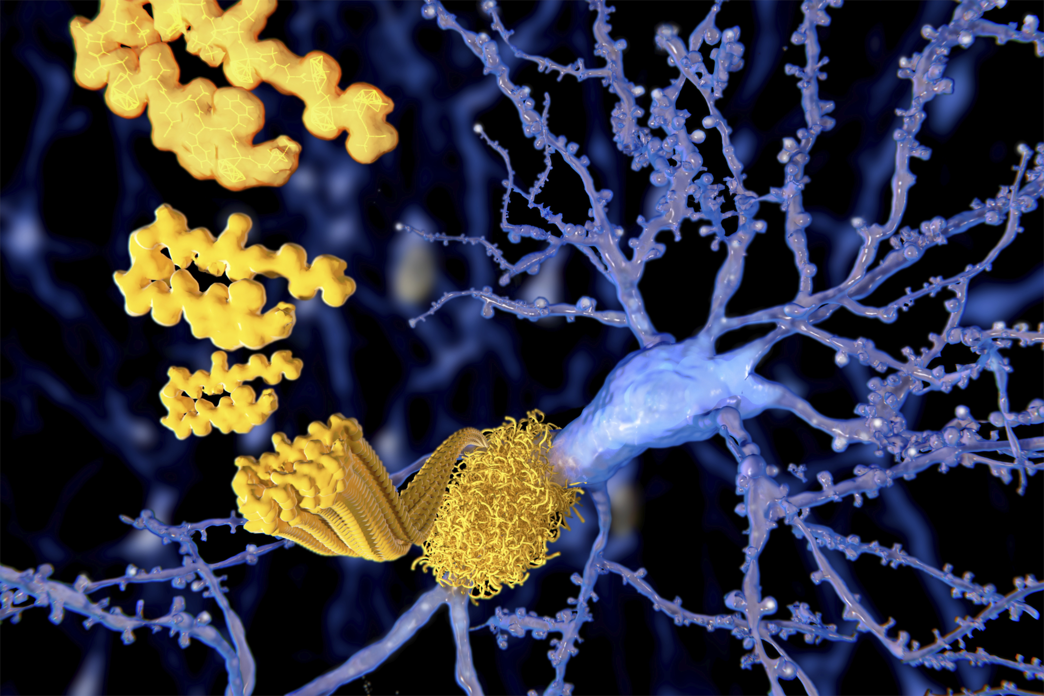
Alzheimer Plaques: fast Visualization in Thick Sections
More than 60% of all diagnosed cases of dementia are attributed to Alzheimer’s disease. Typical of this disease are histological alterations in the brain tissue. So far, there is no cure for this…
Real Time Images of 3D Specimens with Sharp Contrast Free of Haze
THUNDER Imagers deliver in real time images of 3D specimens with sharp contrast, free of the haze or out-of-focus blur typical of widefield systems. They can even image clearly places deep inside a…
Campi di applicazione
Microscopia a fluorescenza
La fluorescenza è uno dei fenomeni fisici più comunemente utilizzati nella microscopia biologica e analitica, soprattutto a causa della sua elevata sensibilità e specificità. Scopri come i microscopi…
Neuroscienze
Stai lavorando per una migliore comprensione delle malattie neurodegenerative o stai studiando la funzionalità del sistema nervoso? Scopri in che modo le soluzioni di imaging di Leica Microsystems…
Ricerca zebrafish
Per ottenere il miglior risultato nella visualizzazione, separazione, manipolazione e imaging è necessario riuscire a osservare i dettagli e le strutture, così da poter prendere la decisione giusta…
Imaging di cellule vive
Spostando la prospettiva dai singoli componenti per microscopio a una soluzione completa, Leica Microsystems integra microscopio, software per l'imaging LAS X, fotocamere e componenti dedicati di…
Ricerca sul cancro
Il cancro è una malattia complessa ed eterogenea causata da cellule che hanno dei difetti nella regolazione della crescita. I cambiamenti genetici ed epigenetici in una cellula o in un gruppo di…
Virology
L’oggetto della tua ricerca si concentra su infezioni e malattie virali? Scopri in che modo puoi approfondire l’aspetto virologico utilizzando le soluzioni di imaging e la preparazione del campione di…
Microscopi per DIC
Un microscopio DIC è basato su un microscopio composto dotato di un filtro di polarizzazione e di un prisma di Wollaston posizionati rispettivamente tra la sorgente luminosa e la lente del…
Microscopio ottico a contrasto di fase
Grazie al microscopio ottico a contrasto di fase è possibile visualizzare le strutture di molti tipi di campioni biologici con maggior contrasto senza la necessità di utilizzare le colorazioni.
Microscopi per campo scuro
Il metodo di contrasto in campo oscuro sfrutta la diffrazione o la dispersione della luce da strutture di un campione biologico o da caratteristiche non uniformi nella struttura di un materiale.

Interessati a saperne di più?
Parlate con i nostri esperti.
Preferite una consulenza personale? Show local contacts