お問い合わせ

暗視野顕微鏡

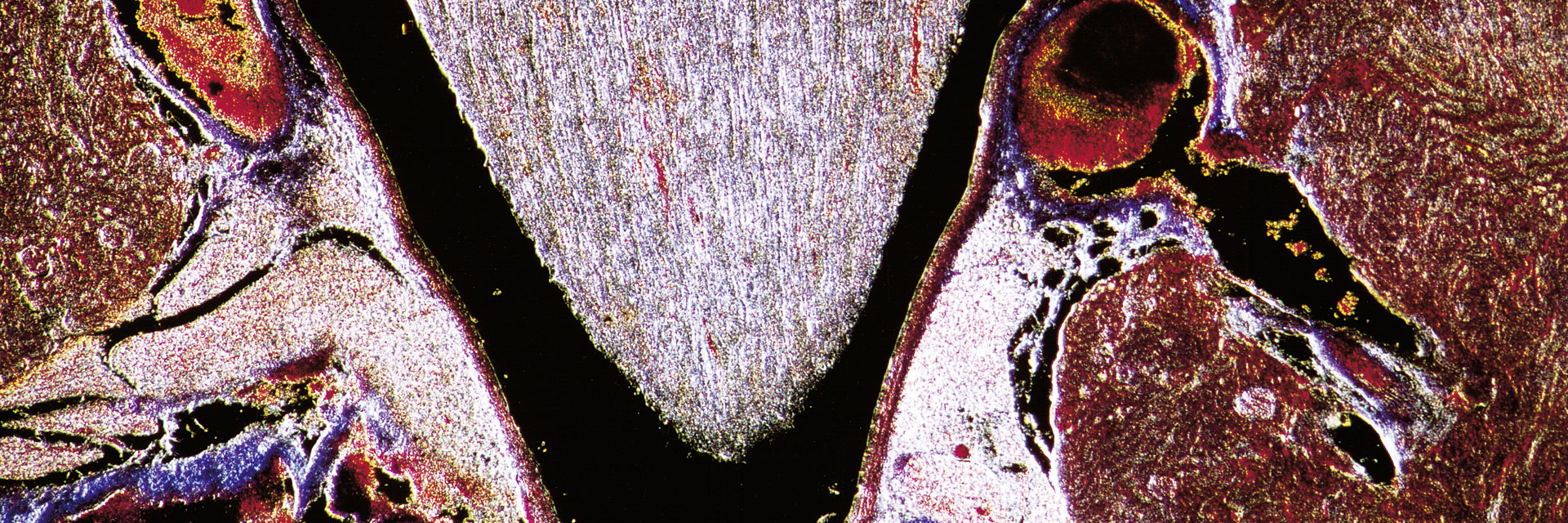
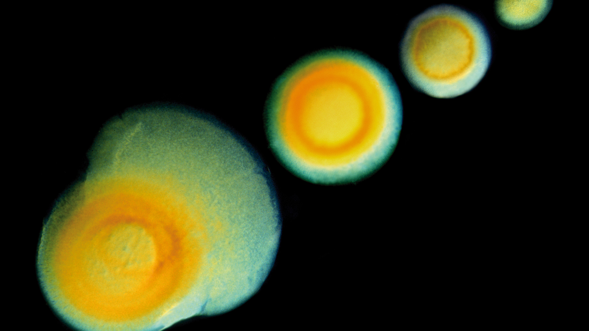
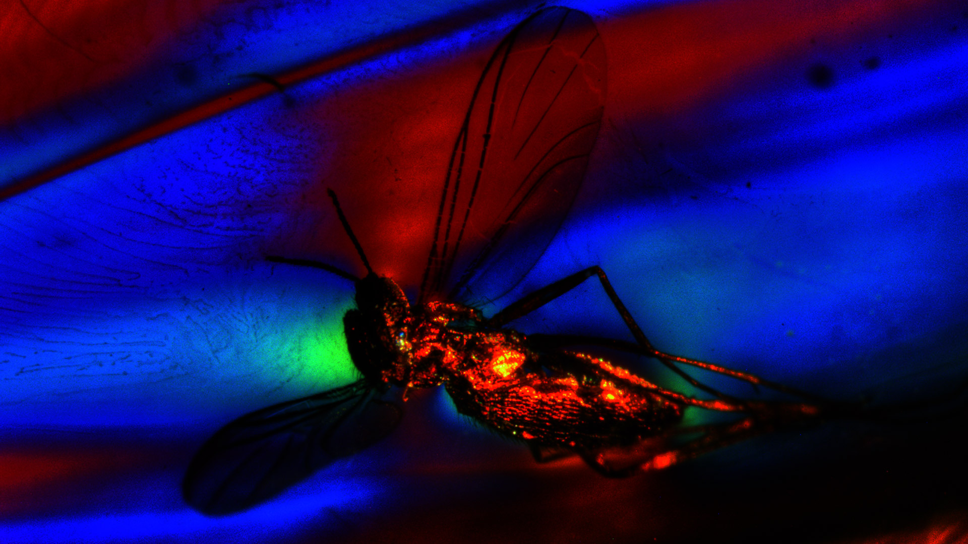
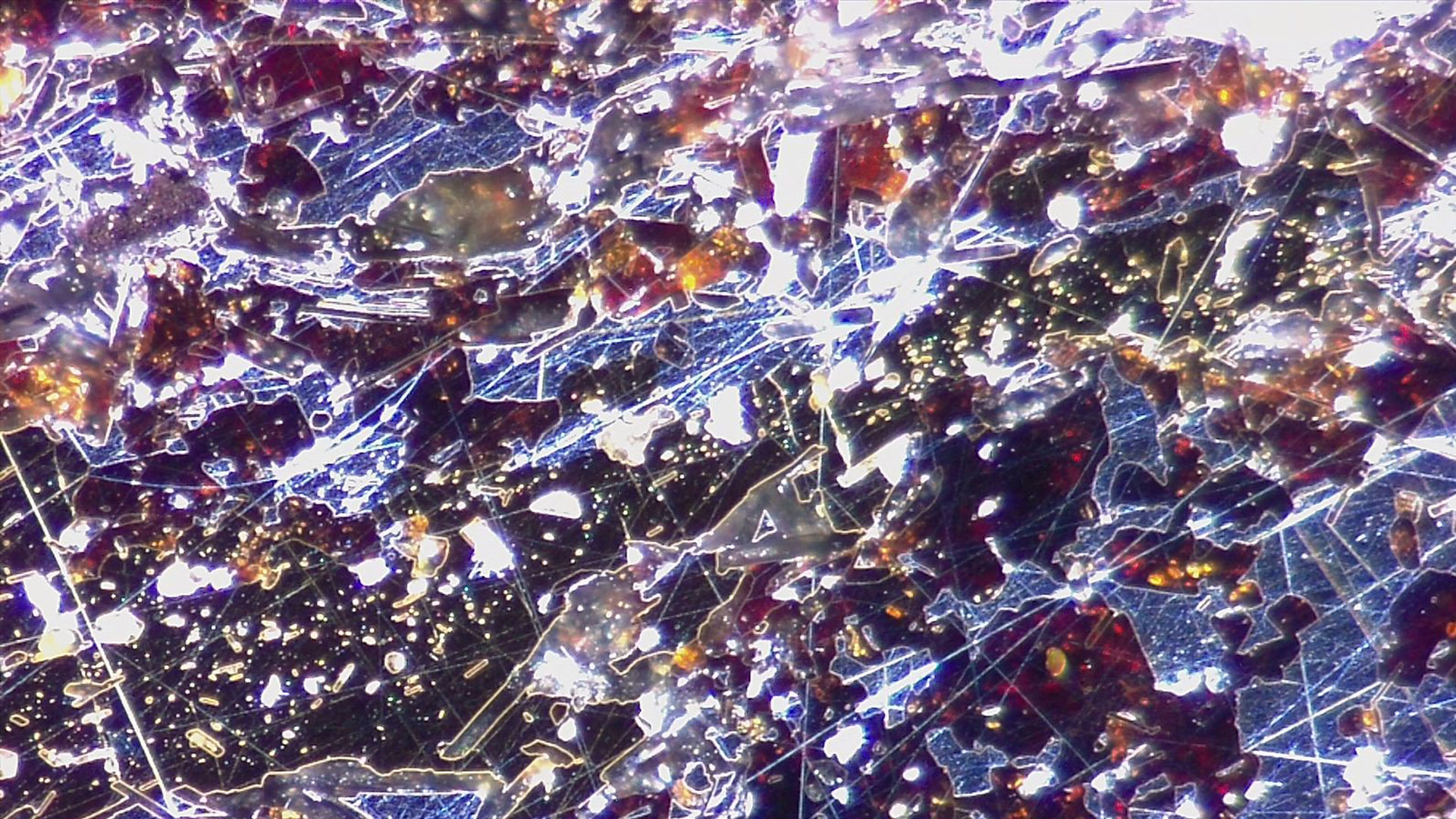
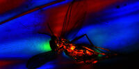
顕微鏡を使用する際、最も一般的なコントラスト法は明視野観察です。 しかし、明視野では通常、多くの透明な生体試料においてコントラストの低い画像しか得られません。 また、多くの透明な物質や反射する不透明な物質の試料でも同様のことが言えます。 このような低コントラストの画像では、細部をほとんど識別することができません。 生物試料の明視野観察では、色素染色を用いてコントラストを高めることができますが、多くの場合、生きた細胞に対して毒性があります。 暗視野顕微鏡では、染色することなく、多くの種類の生物試料の構造を優れたコントラストで観察できます。 また、暗視野顕微鏡は、物質試料を撮影する際にコントラストを高めることができます。 暗視野コントラスト法は、生体試料の構造や物質試料の不均一な特徴から生じる光の回折や散乱を利用する方法です。

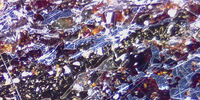
ライカの暗視野顕微鏡は、ライフサイエンスや法医学など様々な用途で、細胞や組織の研究に役立ちます。 暗視野顕微鏡は、物質科学や地球科学の分野でも役立ちます。

お問合せはこちら

お客様のニーズとご予算に合った暗視野照明顕微鏡を専門家がアドバイスいたしますので、ぜひお問い合わせください。

暗視野とは?

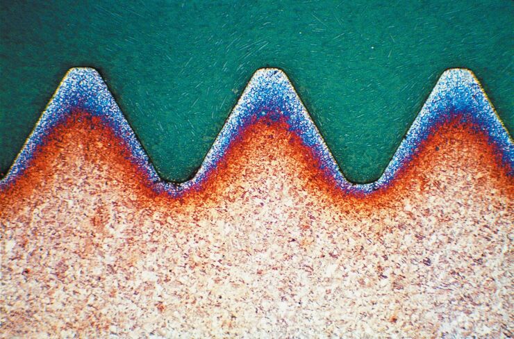
暗視野は、顕微鏡の光学コントラスト技術であり、生物試料の細胞の染色されていない構造を観察できるようにします。 明視野照明では透明に見える細胞構造も、暗視野照明ではより良いコントラストで詳細に見ることができます。 また、透明な物質や不透明な素材の表面にある不均一な特徴は、明視野に比べて、より簡単に暗視野で観察することができます。 細胞内の構造物や物質の特徴は、それらと相互作用する光は散乱し、均一な領域は光を散乱せずに通過するということです。 暗視野顕微鏡を使用すると、細胞の構造や物質の特徴が明るく見え、均一な細胞や物質の背景が暗く見えます。

暗視野顕微鏡の用途は? どういった種類のサンプルを視覚化できるでしょうか?

生体試料や生体組織は、固定されているか生きているかにかかわらず、暗視野顕微鏡で観察されることが多くあります。 また、暗視野顕微鏡では、多種多様な物質試料や地質試料も観察できます。 たとえば、次の記事をご覧ください。 "実体顕微鏡で発生生物学をより効率的に学ぶ: ゼブラフィッシュ、メダカ、アフリカツメガエル""金属合金の粒度分布測定をニーズに合わせて行うには?"

暗視野顕微鏡の仕組み

暗視野顕微鏡は、光源と集光レンズの間に環状の絞りを用いた複合顕微鏡です。 

絞りを通過したリング状の光は、コンデンサーによって観察したい生体試料や物質試料に集光されます。 リング状の光の一部が、試料の構造や特徴によって回折または散乱します。 回折した光が対物レンズに集光されます。 一方、リング状の光のうち、試料をそのまま通過する部分や、散乱せずに試料で反射する部分は、対物レンズで集光されません。 試料の構造や特徴によって散乱した光は、光の散乱がない試料の背景部分よりも明るく見えるようになります。

詳しくは、こちらの記事をご覧ください: "メタログラフィー - 入門編"

ライフサイエンス研究

ライカの暗視野顕微鏡は、ライフサイエンス研究において、生物学的構造や細胞プロセスの可視化、分析、文書化によく使用されています。

Read More

法医学

ライカの暗視野顕微鏡は、塗料、顔料、繊維、人体組織など、法医学における証拠調べに、非常に有効なソリューションです。

Read More

マテリアルサイエンスと地球惑星

ライカの暗視野顕微鏡は、透明・不透明な物質、鉱物、結晶、高分子の研究に大きな威力を発揮します。

Read More

よくある質問 暗視野顕微鏡

Show answer 明視野顕微鏡と暗視野顕微鏡の違いは何ですか?

暗視野顕微鏡は、光源の前に環状の絞りを用いる以外は、従来の明視野顕微鏡と同じです。 暗視野照明からの光は高い入射角で試料に入射し、試料を透過するかまたはその表面で反射します。対物レンズを通過した光は、最後に接眼レンズを通過またはカメラセンサーに到達します。 暗視野照明は、透明な試料の均一な領域や不透明な試料の均一な表面が暗く見えます。これは、高い入射角の光の大部分が対物レンズには入らないためです。 通常、透明な試料や不透明な試料の表面は、光を散乱させるという特徴があります。 そのため、暗視野顕微鏡の画像では、これらの特徴により散乱した光が対物レンズに入射するため、暗い背景とこれらの特徴に対応する明るい領域が表示されます。

Show answer 暗視野顕微鏡に生きた試料を使用することはできますか?

はい、細胞や生物などの生きた生物試料は、暗視野顕微鏡で簡単に観察することができます。

Show answer 暗視野顕微鏡にカメラなどのアクセサリーを接続して使用することはできますか?

はい、暗視野顕微鏡には、コントラスト法で観察した画像を記録するためのカメラを搭載することができます。 また、他のアクセサリーを取り付けることも可能です。 詳細については、ライカの担当者にお問い合わせください。

関連記事

暗視野顕微鏡に関する最新記事を読む

ライカ マイクロシステムズのサイエンスラボでは、 顕微鏡に関する専門記事やお客様事例、基本情報を提供しています。 日常業務や研究で、ビギナーから経験豊富な方まで幅広くサポートします。

その他の記事
Images of the same area of a processed wafer taken with standard (left) 
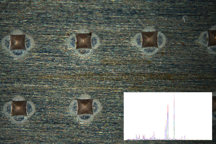
and oblique (right) brightfield illumination using a Leica compound microscope. The defect on the wafer surface is clearly more visible with oblique illumination.

Rapid Semiconductor Inspection with Microscope Contrast Methods

Semiconductor inspection for QC of materials like wafers can be challenging. Microscope solutions that offer several contrast methods enable fast and reliable defect detection and efficient workflows.

Fast Visual and Chemical Analysis of Contamination and Underlying Layers

Visual and chemical analysis of contamination on materials with a 2-methods-in-1 solution leading to an efficient, more complete analysis workflow is described in this report. A 2-in-1 solution,…

Metallography – an Introduction

This article gives an overview of metallography and metallic alloy characterization. Different microscopy techniques are used to study the alloy microstructure, i.e., microscale structure of grains,…
Measuring grains size with Abrams Three-Circle Procedure.

How to Adapt Grain Size Analysis of Metallic Alloys to Your Needs

Metallic alloys, such as steel and aluminum, have an important role in a variety of industries, including automotive and transportation. In this report, the importance of grain size analysis for alloy…
Transgenic zebrafish larva where fluorescent proteins label the heart muscle blue, blood and blood vessels red, and all circulatory system cells green. Image recorded with a M205 FA microscope.

Imaging and Analyzing Zebrafish, Medaka, and Xenopus

Discover how to image and analyze zebrafish, medaka, and Xenopus frog model organisms efficiently with a microscope for developmental biology applications from this article.
C. elegans

Studying Caenorhabditis elegans (C. elegans)

Find out how you can image and study C. elegans roundworm model organisms efficiently with a microscope for developmental biology applications from this article.

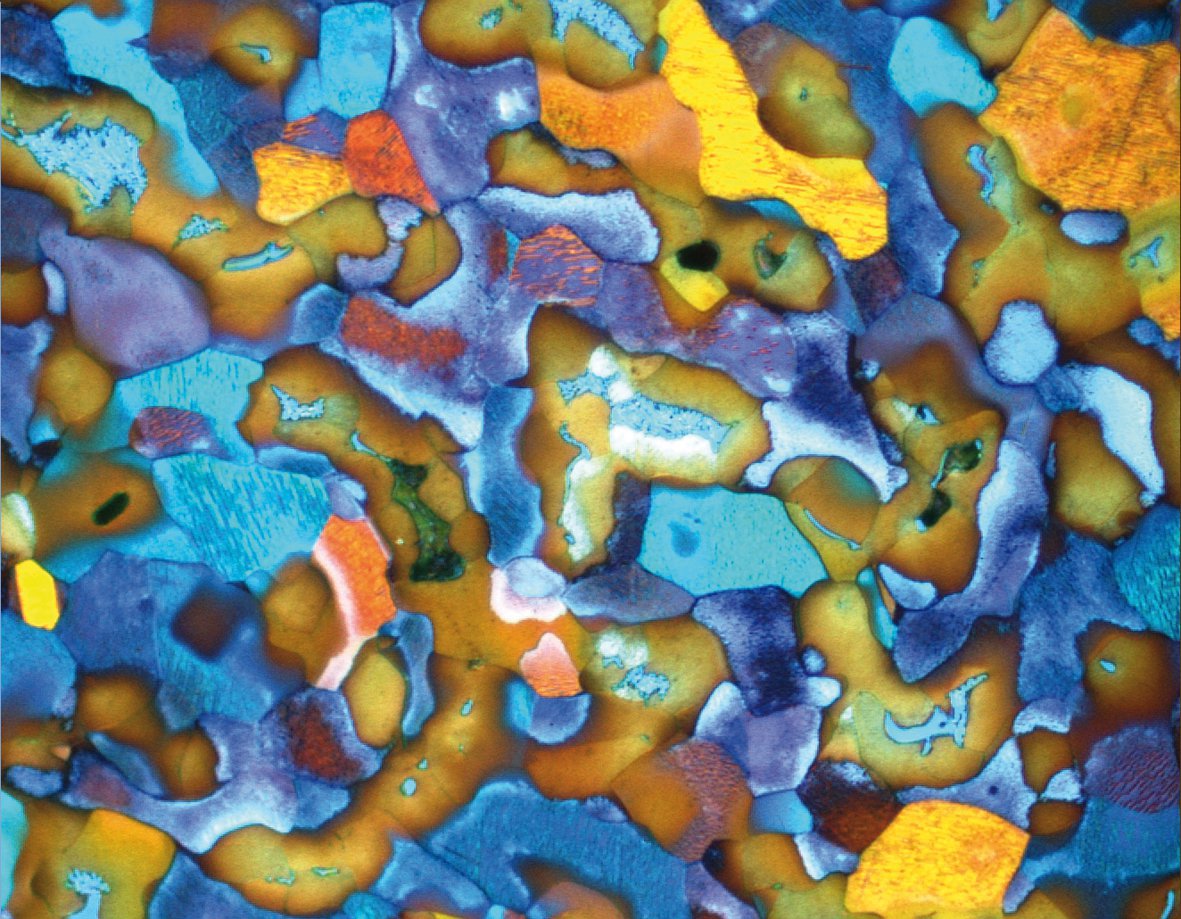
Metallography with Color and Contrast

The examination of microstructure morphology plays a decisive role in materials science and failure analysis. There are many possibilities of visualizing the real structures of materials in the light…
Section taste buds rabbit, differential interference contrast microscope

Optical Contrast Methods

Optical contrast methods give the potential to easily examine living and colorless specimens. Different microscopic techniques aim to change phase shifts caused by the interaction of light with the…
Background image
Scroll to top