Mica
제품소개
홈
Leica Microsystems
Mica 세계 최초의 이미징 Microhub를 만나보세요.
이는 모든 것을 변화시킵니다.
최신 기사를 읽어 보세요

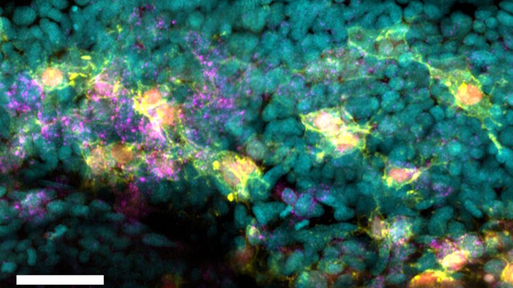
How do Cells Talk to Each Other During Neurodevelopment?
Professor Silvia Capello presents her group’s research on cellular crosstalk in neurodevelopmental disorders, using models such as cerebral organoids and assembloids.

How to Streamline Your Histology Workflows
Streamline your histology workflows. The unique Fluosync detection method embedded into Mica enables high-res RGB color imaging in one shot.
 imaged with the THUNDER Imager 3D Cell Culture. Courtesy of Dr. F.T. Arroso Martins, Tamere University, Finland.")
How to Get Deeper Insights into your Organoid and Spheroid Models
In this eBook, learn about key considerations for imaging 3D cultures, such as organoids and spheroids, and discover microscopy solutions to shed new insights into dynamic processes in 3D real-time

Epi-Illumination Fluorescence and Reflection-Contrast Microscopy
This article discusses the development of epi-illumination and reflection contrast for fluorescence microscopy concerning life-science applications. Much was done by the Ploem research group…
")
Introduction to Fluorescent Proteins
Overview of fluorescent proteins (FPs) from, red (RFP) to green (GFP) and blue (BFP), with a table showing their relevant spectral characteristics.

Examining Developmental Processes In Cancer Organoids
Interview: Prof. Bausch and Dr. Pastucha, Technical University of Munich, discuss using microscopy to study development of organoids, stem cells, and other relevant disease models for biomedical…
![[Translate to korean:] Masson-Goldner staining of a hedgehog brain slice.](/fileadmin/_processed_/c/0/csm_Masson-Goldner_staining_of_hedgehog_brain_slice_dd9e8977dd.jpg "[Translate to korean:] Masson-Goldner staining of a hedgehog brain slice.")
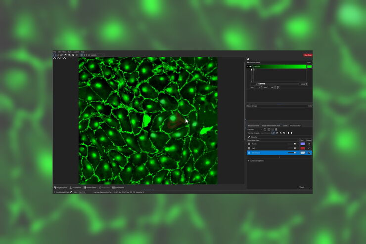
How to Image Histological and Fluorescent Samples with One System
VIDEO ON DEMAND - How to image histological and fluorescent samples with one system. FluoSync, the new technology embedded into Mica enables the imaging of both histological staining and fluorescence…
How to Radically Simplify Workflows in Your Imaging Facility
VIDEO ON DEMAND - How to radically simplify imaging workflows and generate meaningful results with less time and effort using a highly automated microscope that unites widefield and confocal imaging.
FluoSync - a Fast & Gentle Method for Unmixing Multicolour Images
In this white paper, we focus on a fast and reliable method for obtaining high-quality multiplex images in fluorescence microscopy. FluoSync combines an existing method for hybrid unmixing with…
Harnessing Microfluidics to Maintain Cell Health During Live-Cell Imaging
VIDEO ON DEMAND - In this episode of MicaCam, we will use microfluidics to explore the effect of shear stress on cell morphology, examine the effect of nutrient replenishment on cellular growth during…
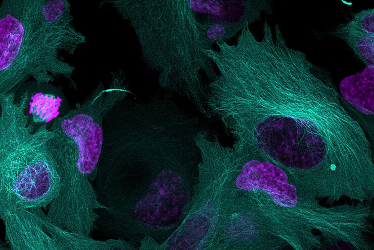
, SPY-Actin (cyan), and SiR-Tubulin (magenta). Instant Computational Clearing (ICC) was applied.")
How to Perform Dynamic Multicolor Time-Lapse Imaging
Live-cell imaging sheds light on diverse cellular events. As many of these events have fast dynamics, the microscope imaging system must be fast enough to record every detail. One major advantage of…
 and CellEvent™ (yellow).")
Following Multiple Events during Staurosporine Apoptosis
Coming next on MicaCam - Livestream on 19th October 2022 - In this episode of MicaCam, we show how adding additional markers to an apoptosis kit can markedly increase the amount of information a…
, the cis-golgi matrix protein GM130 (AF488, green), and the trans-golgi network membrane protein TGN46 (AF647, red).")
Golgi Organizational Changes in Response to Cell Stress
VIDEO ON DEMAND - In this episode of MicaCam, our special guest George Galea from EMBL Heidelberg will look at HeLa Kyoto cells treated with various chemotherapeutic agents to investigate their effect…
 stained to show the nucleus")
AI-Enabled Spatial Analysis of Complex 3D Datasets
VIDEO ON DEMAND - This edition of MicaCam offers practical advice on the extraction of publication grade insights from microscopy images. Our special guest Luciano Lucas (Leica Microsystems) will…

3D Tissue Imaging: From Fast Overview To High Resolution With One Click
3D Tissue imaging is a widespread discipline in the life sciences. Researchers use it to reveal detailed information of tissue composition and integrity, to make conclusions from experimental…

How To Perform Fast & Stable Multicolor Live-Cell Imaging
With the help of live-cell imaging researchers gain insights into dynamic processes of living cells up to whole organisms. This includes intracellular as well as intercellular activities. Protein or…

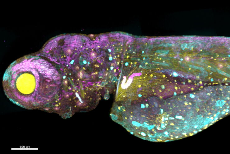
Imaging of Cardiac Tissue Regeneration in Zebrafish
Learn how to image cardiac tissue regeneration in zebrafish focusing on cell proliferation and response during recovery. MicaCam Episode 04 with Laura Peces-Barba Castaño from the Max Planck…
How Does The Cytoskeleton Transport Molecules?
VIDEO ON DEMAND - See how 3D cysts derived from MDCK cells help scientists understand how proteins are transported and recycled in tissues and the role of the cytoskeleton in this transport.
 embryo, from sphere stage to somite stages.")
Studying Early Phase Development of Zebrafish Embryos
VIDEO ON DEMAND - This second edition of MicaCam focuses on combining widefield and confocal imaging to study the early-stage development of zebrafish embryos (Danio rerio), from oocyte to…

Multi-Color Caspase 3/7 Assays with Mica
Caspases are involved in apoptosis and can be utilized to determine if cells are undergoing this programmed cell death pathway in so-called caspase assays. These assays can be run by e.g. flow…

How To Get Multi Label Experiment Data With Full Spatiotemporal Correlation
VIDEO ON DEMAND - The first edition of MicaCam focuses on the special challenges of live cell experiments. Our hosts Lynne Turnbull and Oliver Schlicker use the example of studying the mitochondrial…

Simplifying Complex Fluorescence Multiwell Plate Assays
Apoptosis, or programmed cell death, occurs during organism embryo development to eliminate unwanted cells and during healing in adults to rid the body of damaged cells and help prevent cancer.…

Efficient Long-term Time-lapse Microscopy
When doing time-lapse microscopy experiments with spheroids, there are certain challenges which can arise. As the experiments can last for several days, prolonged sample survival must be achieved…
How to Prepare your Specimen for Immunofluorescence Microscopy
Immunofluorescence (IF) is a powerful method for visualizing intracellular processes, conditions and structures. IF preparations can be analyzed by various microscopy techniques (e.g. CLSM,…
Live-Cell Imaging Techniques
The understanding of complex and/or fast cellular dynamics is an important step for exploring biological processes. Therefore, today’s life science research is increasingly focused on dynamic…
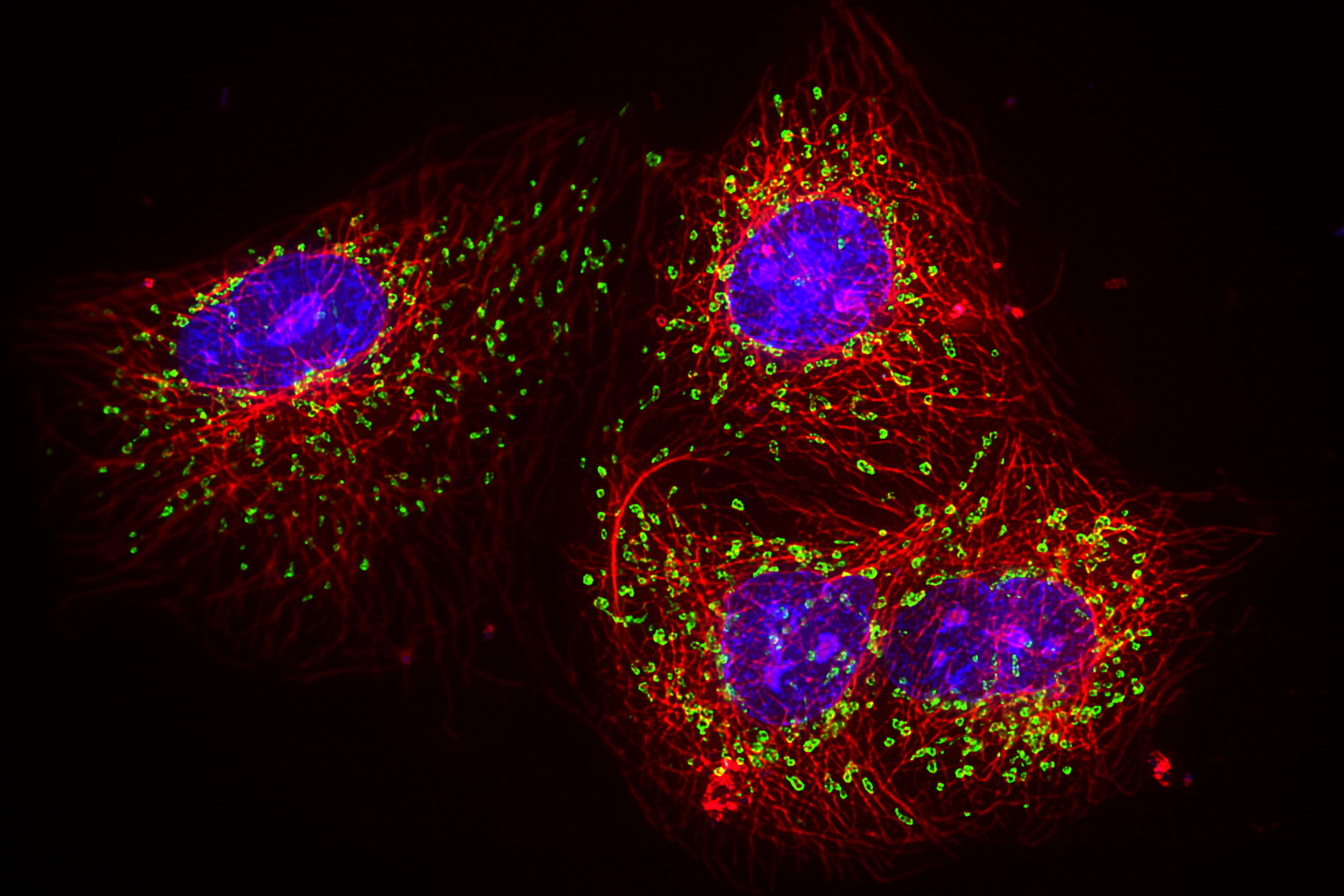
Fluorescent Dyes
A basic principle in fluorescence microscopy is the highly specific visualization of cellular components with the help of a fluorescent agent. This can be a fluorescent protein – for example GFP –…
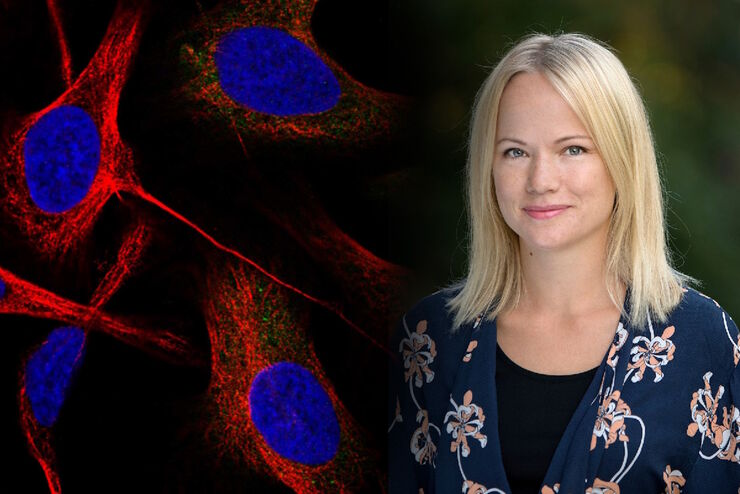
Applying AI and Machine Learning in Microscopy and Image Analysis
Prof. Emma Lundberg is a professor in cell biology proteomics at KTH Royal Institute of Technology, Sweden. She is also the director of the Cell Atlas, an integral part of the Swedish-based Human…
Using Machine Learning in Microscopy Image Analysis
Recent exciting advances in microscopy technologies have led to exponential growth in quality and quantity of image data captured in biomedical research. However, analyzing large and increasingly…
The AI-Powered Pixel Classifier
Achieving reproducible results manually requires expertise and is tedious work. But now there is a way to overcome these challenges by speeding up this analysis to extract the real value of the image…
, Astrocytes (GFAP, red), Nuclei (DAPI, blue).")
Multicolor Microscopy: The Importance of Multiplexing
The term multiplexing refers to the use of multiple fluorescent dyes to examine various elements within a sample. Multiplexing allows related components and processes to be observed in parallel,…
Considerations for Multiplex Live Cell Imaging
Simultaneous multicolor imaging for successful experiments: Live-cell imaging experiments are key to understand dynamic processes. They allow us to visually record cells in their living state, without…
A New Method for Convenient and Efficient Multicolor Imaging
The technique combining hyperspectral unmixing and phasor analysis was developed to simplify the process of getting images from a sample labeled with multiple fluorophores. This aggregate method…
 and astrocytes (green) in a cortical spheroid derived from human induced pluripotent stem cells.")
Neuroscience Images
Neuroscience commonly uses microscopy to study the nervous system’s function and understand neurodegenerative diseases.

Introduction to Widefield Microscopy
This article gives an introduction to widefield microscopy, one of the most basic and commonly used microscopy techniques. It also shows the basic differences between widefield and confocal…
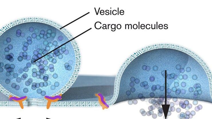
Nobel Prize 2013 in Physiology or Medicine for Discoveries of the Machinery Regulating Vesicle Traffic
On October 7th 2013, The Nobel Assembly at Karolinska Institutet has decided to award The Nobel Prize in Physiology or Medicine 2012 jointly to James E. Rothman, Randy W. Schekman and Thomas C. Südhof…

Nobel Prize 2012 in Physiology or Medicine for Stem Cell Research
The Nobel Prize recognizes two scientists who discovered that mature, specialised cells can be reprogrammed to become immature cells capable of developing into all tissues of the body. Their findings…
 inoculated with cowpea mosaic virus (CPMV) containing the GFP-gene inserted between the movement protein (MP) and the capsid proteins (CPs) in the viral RNA 2")
Introduction to Live-Cell Imaging
The understanding of complex and fast cellular dynamics is an important step to get insight into biological processes. Therefore, today’s life science research more and more demands studying…

An Introduction to Fluorescence
This article gives an introduction to fluorescence and photoluminescence, which includes phosphorescence, explains the basic theory behind them, and how fluorescence is used for microscopy.
적용 분야
형광 현미경법
형광은 주로 높은 감도와 높은 특이성 때문에 생물학적 및 분석적 현미경법에서 가장 일반적으로 사용되는 물리적 현상 중 하나입니다. 연구에 형광 현미경이 어떻게 활용될 수 있는지 확인해보세요.
세포생물학
인간의 건강과 질병을 기준으로 세포를 이해하는 것에 연구의 초점이 맞추어져 있다면 관심 세포를 시공간 및 분자 측면에서 자세히 조사하는 것은 매우 중요합니다. 이는 현미경이 세포생물학에서 매우 중요한 도구인 이유입니다. 현미경을 사용하면 세포 기관과 고분자를 분석할 뿐만 아니라, 시료의 구조적 환경 내에서 시료를 자세히 연구할 수 있습니다. 세포생물학…
암 연구
암은 성장 통제에 결함이 있는 세포에 의해 발생하는 복잡하고 이질적인 질병입니다. 하나 또는 한 그룹의 세포에서 일어나는 유전적 또는 후생적 변화가 정상적인 기능을 방해하고, 자율적이고 통제되지 않는 세포 성장과 증식을 초래합니다.
라이브 셀 이미징
Leica Microsystems는 단일 현미경 구성 요소에서 완전한 라이브 셀 이미징 솔루션으로 관점을 전환해 현미경, LAS X 이미징 소프트웨어, 카메라 및 전용 타사 구성요소를 완전한 라이브 셀 이미징 시스템에 통합합니다.
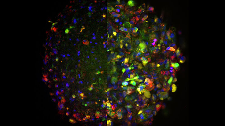
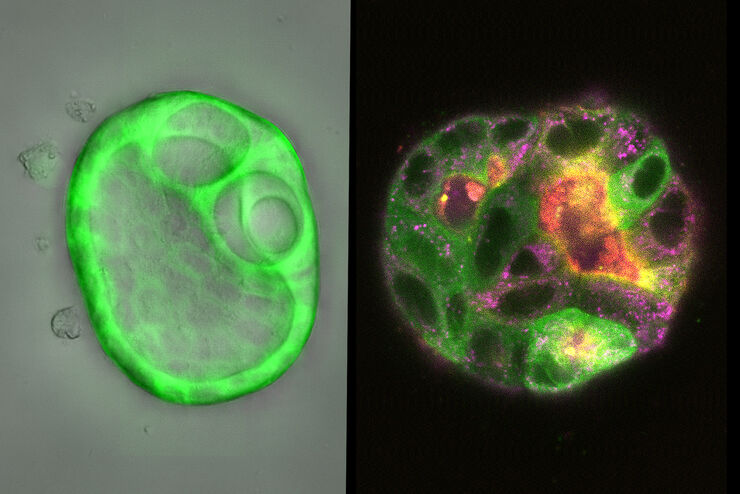
오가노이드와 3D 세포 배양
최근 생명과학 연구에서 가장 흥미로운 발전 중 하나는 오가노이드, 스페로이드 또는 장기 칩 모델과 같은 3D 세포 배양 시스템의 개발입니다. 3D 세포 배양이란 세포가 3차원에서 성장하고 주변 환경과 상호작용할 수 있는 인위적인 환경입니다. 이러한 조건은 체내 상태와 유사합니다.
연구 분야의 모델 유기체
모델 유기체는 연구자들이 특정한 생물학적 과정을 연구하기 위해 사용하는 종입니다. 이들은 인간과 유사한 유전적 특성을 가지고 있으며, 유전학, 발달생물학, 신경과학 같은 연구 분야에서 일반적으로 사용됩니다. 유기체 모델은 일반적으로 실험실 환경에서 쉬운 유지와 번식, 짧은 세대 주기 또는 특정 형질이나 질병을 연구하기 위한 돌연변이 생성 능력 때문에…
신경과학 연구
신경변경 질환에 대해 더 잘 이해하기 위해 노력하고 있거나 신경계 기능을 연구하고 계십니까? 라이카마이크로시스템즈의 이미지 솔루션을 통해 발전을 이룰 수 있는 방법을 알아보세요.

더 자세히 알고 싶으신가요?
전문가와 상담하세요.
개인 컨설팅을 원하십니까? Show local contacts