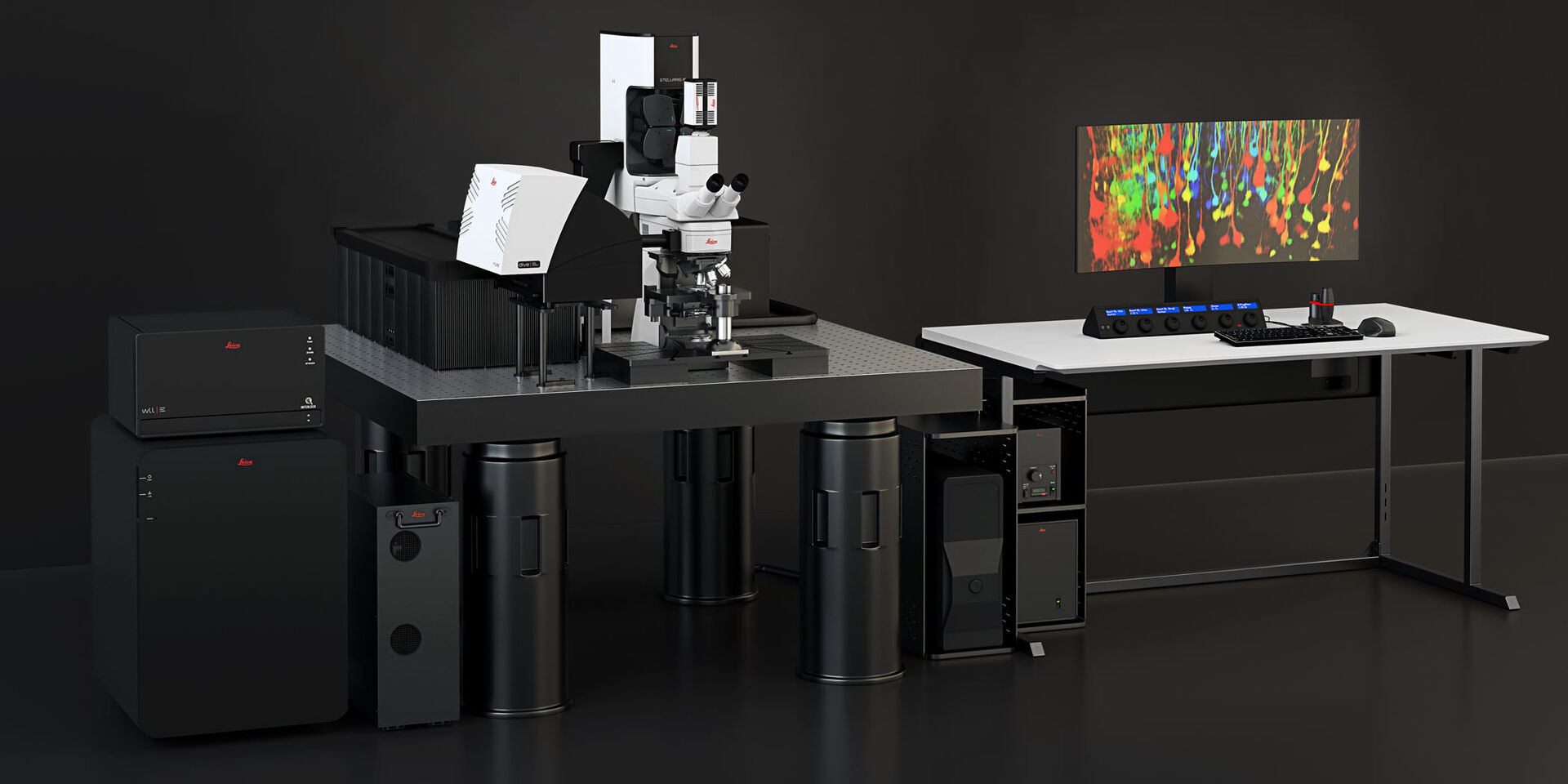
STELLARIS 8 DIVE Multiphoton Microscope
Multicolor Multiphoton gets you closer to the truth
The combination of multiple distinct fluorescent labels is increasingly used to study dynamic interaction and spatial relationships among cells and molecules. The goal is to understand a myriad of complex biological events, such as cellular connectivity, cell phenotyping, protein interaction or co-expression, and co-localization. Scaling up such studies to whole organs or tissues requires suitable large-volume multicolor microscopy methods. DIVE and STELLARIS have now merged to provide you with the power of flexible multicolor multiphoton imaging. Furthermore, you can expand the potential of your experiments with additional label-free imaging capabilities.
Seamlessly integrated into the confocal software interface, STELLARIS 8 DIVE multiphoton microscope offers the benefits of high speed and excellent navigation so that dynamic processes in complex samples can be studied with ease. STELLARIS 8 DIVE – A rainbow of possibilities for your research.
The power to analyze in vivo with more detail than ever before
The STELLARIS 8 DIVE multiphoton microscope provides you with flexible multicolor imaging beyond 1 mm in depth Thanks to 4Tune, a spectrally tunable non-descanned detector, you can define up to four detection bands simultaneously or an unlimited number when imaged sequentially anywhere in the emission spectrum It gives you the flexibility to adapt to the combination of fluorophores you need. With STELLARIS 8 DIVE, you can perform multiphoton experiments with more than a billion possible fluorophore combinations, empowering you to study complex processes, such as neuronal connectivity, organ structure, dynamic interactions, or spatial relationships of cells and proteins in much more detail.
Study metastasis in live specimens using four or more colors to distinguish relevant proteins, hippocampal activity in awake mice, or the structure of fixed thick intestine sections with STELLARIS 8 DIVE!
The conventional dichroics are never optimal to distinguish all fluorophores, but with the spectral detectors, this is now possible and much easier, since we can really optimize for each fluorophore the wavelengths you want to detect.
Prof. Dr. Jacco van Rheenen. Netherlands Cancer Institute, Amsterdam (the Netherlands).

DIVE with ease – the 4Tune detector
The 4Tune non-descanned detection system can be equipped with 2 to 4 detectors and is freely configurable with hybrid detectors (Power HyD NDD), photomultipliers (PMTs), or a mix of both. The emission light is separated by a combination of variable dichroics and bandpass filters. Freely tune your detection over the whole visible spectrum (380 – 800 nm)!
The 4Tune user interface allows you to optimize the emission setting for multiple transgenic markers by simple drag and drop. Due to its clear and intuitive design, operating it is easy and requires minimal training.
With the STELLARIS 8 DIVE, you are equipped for every existing and newly developed transgenic marker and future proved for new developments!

Explore new dimensions in depth
With the STELLARIS 8 DIVE multiphoton microscope, you can tune for the deepest insight and finest detail. All excitation beams can be optimally adjusted independently for any objective using the new Vario Beam Expander (VBE).
The VBE allows for optimized colocalization and the right balance of resolution and depth in line with your research question.

Optimize for depth and resolution with the Vario Beam Expander
The Leica Vario Beam Expander VBE combines tunable beam diameter and tunable divergence. This offers you maximum depth, best resolution and full color correction.
Tunable beam diameter for best balance of resolution and depth
The STELLARIS 8 DIVE allows you to adapt to your sample requirements. Using the Vario Beam Expander, you can choose: Maximum resolution – which results from a fully illuminated back aperture of your objective lens – and maximized penetration depth – resulting from slightly underfilling the back aperture. Underfilling the back-aperture results in a larger focal volume and a reduced pathlength which leads to more efficient excitation.
Tunable beam divergence for full color correction
Our IR APO objectives do not suffer from chromatic aberations over the IR range. However, with STELLARIS 8 DIVE you are ready to use objective lens suitable for IR along with multiple IR laser lines: The Vario Beam Expander can be used to correct for chromatic shifts and enable meaningful multicolor experiments.

Expand the potential of deep in vivo experiments with label-free imaging
Molecules such as collagen and elastin have relevant roles in diseases like cancer. Our 4-tune detector enables the use of second and third harmonic generation signals that allow you to study these important structures without staining.
The combination of DIVE with STELLARIS also enables the use of lifetime-based information intrinsic to fluorescence. This ability allows you to perform experiments like metabolic mapping of a specimen via lifetime imaging of NADH or FAD.

Navigate tissue easily without the need of additional staining
Navigating through tissue often requires orientation landmarks to know where areas of interest are located. The scaffolding property of collagen can help in navigating through tissue and find areas of interest without the need of a counterstain.
Most biological tissues contain collagen which is the main component of connective tissues. For example, intestines are surrounded by a layer of collagen. Collagen can be visualized with multiphoton microscopy easily by collecting emission signals at exactly ½ of the excitation wavelength. With the flexible detection windows in 4Tune, any wavelength can be used to collect this signal, so no additional label or effort is needed.
Combining multiphoton imaging and lifetime information to study metabolic changes
Metabolic changes can be an important marker for the health of tissue.
STELLARIS 8 DIVE offers you all the benefits of TauSense, a set of imaging tools based on fluoresences lifetime. When the metabolic state of a cell changes, it can be visualized by changes in the fluorescence lifetime of molecules, such as NADH. NADH plays a main role in the metabolism of sugar and its fluorescence lifetime is dependent on the glucose concentration. The NADH fluorescence lifetime is altered by a conformational change that occurs due to the biochemical reaction causing the breakdown of glucose.
For full quantitative fluorescence lifetime analysis, STELLARIS 8 DIVE can be combined with FAst Lifetime COntrast (FALCON).

Add additional dimensions to your multiphoton microscopy experiments
Autofluorescence is a natural fluorescence emission from tissues arising from endogenous fluorophores, such as small molecules like NADH or FAD, or tissue structures. It often creates a problem when imaging specimens. But what if you could use it to your advantage?
Thanks to the combination of DIVE and TauSense, you can now use lifetime-based separation to obtain valuable information from autofluorescence signals. This capability gives you an additional channel enabling you to get more information from your valuable specimens.

Gain productivity with STELLARIS’ unique software capabilities
Multiphoton microscopes are usually rigid to use and need to be adapted to each experiment and user. Add to this the stress of working with live animals or freshly explanted tissue and you quickly understand the advantage of having flexibility when doing multiphoton experiments. STELLARIS 8 DIVE provides you with an easy, hassle-free workflow from setup to final results thanks to the seamless integration of multiphoton capabilities into the STELLARIS software.
- Seamless experimental setup with ImageCompass
- Intuitive approach for finding an area of interest on your sample with LAS X Navigator
- Boosting of both speed and resolution with Dynamic Signal Enhancement

Easy and fast setup of multicolor multiphoton imaging with ImageCompass
STELLARIS 8 DIVE multiphoton hardware is fully integrated into the ImageCompass interface of STELLARIS, allowing you to easily define your experimental settings for a quick start.
MP excitation and emission can be automatically defined by the system using the extensive database of fluorophores. They can also be defined manually with only a few clicks. Sequential settings and a fast live, 3D viewer – multicolor multiphoton imaging was never as easy.

Explore relevant details instantly while always keeping an overview of your specimen
The LAS X Navigator is a powerful navigation tool that enables you to quickly switch from searching image by image to seeing a full overview of your sample. Thanks to the integration of DIVE and STELLARIS, your multiphoton experiments can become more efficient. Benefit from the ability to freely navigate through your large and complex samples, providing deep multicolor imaging with fast overviews, multi-position imaging, and tile scans.
A mosaic image of a kidney slice, 1 cm long and 0.5 mm thick, is easily acquired and gives a full picture of the kidney nerve cell and collagen system (here in combination with TauContrast).


Dynamic Signal Enhancement:
Maintain resolution of fast processes in vivo
Processes in live specimens can be fast. Fluorescent Signals in animals models, however, tend to be weak.
The solution to account for both challenges is Dynamic Signal Enhancement. It allows averaging for better S/N and, as consequence, better resolution, while adapting to your sample’s dynamics.

Interested to know more?
Talk to our experts.
Do you prefer personal consulting? Show local contacts