STELLARIS DLS
Confocal Microscopes
Products
Home
Leica Microsystems
STELLARIS DLS Digital Light Sheet Microscope
Light Sheet Re-Imagined
Read our latest articles

Notable AI-based Solutions for Phenotypic Drug Screening
Learn about notable optical microscope solutions for phenotypic drug screening using 3D-cell culture, both planning and execution, from this free, on-demand webinar.
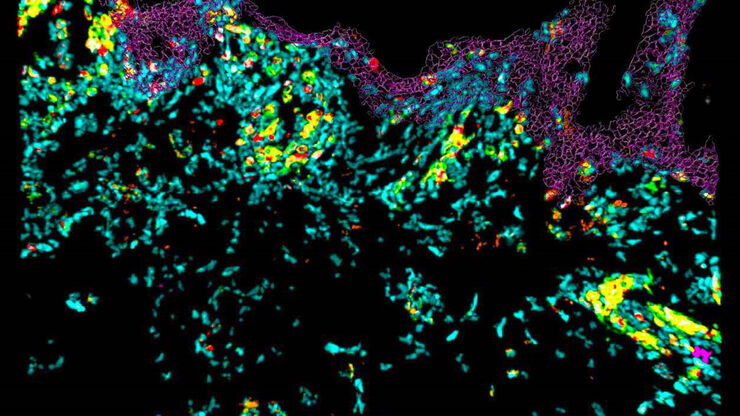
Confocal Imaging of Immune Cells in Tissue Samples
In this webinar, you will discover how to perform 10-color acquisition using a confocal microscope. The challenges of imaged-based approaches to identify skin immune cells. A new pipeline to assess…

Virtual Reality Showcase for STELLARIS Confocal Microscopy Platform
In this webinar, you will discover how to perform 10-color acquisition using a confocal microscope. The challenges of imaged-based approaches to identify skin immune cells. A new pipeline to assess…
")
Wt1 Genes Can Induce a Cardiomyocyte to Epicardial-like Cell Fate Transition
From this study, it was concluded that Wt1 plays a yet undescribed role for cardiomyocyte differentiation by repressing chromatin opening at specific genomic loci and that sustained ectopic expression…

Gentle and Flexible 3D Imaging of Biological and Cleared Samples
For 3D imaging of biological samples, light sheet microscopy is a powerful and sensitive tool allowing you to measure (sub)cellular dynamics over long periods of time. Meanwhile, confocal microscopy,…
 and mito OM (red) in a live U2OS cell")
Multicolor 4D Super Resolution Light Sheet Microscopy
The AI Microscopy Symposium offers a unique forum for discussing the latest AI-based technologies and tools in the field of microscopy and biomedical imaging. In this scientific presentation, Yuxuan…

Imaging of Anti-Cancer Drug Uptake in Spheroids using DLS
Spheroid 3D cell culture models mimic the physiology and functions of living tissues making them a useful tool to study tumor morphology and screen anti-cancer drugs. The drug AZD2014 is a recognized…
Effects of Clearing Media on Tissue Transparency and Shrinkage
This study comprehensively evaluates the effects of different clearing media on tissue transparency and shrinkage by comparing freshly dissected dipteran fly brains with their cleared equivalents.…
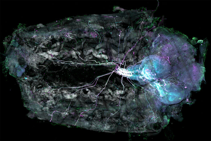
Understanding Motor Sequence Generation Across Spatiotemporal Scales
We have developed a microscopy-based pipeline to characterize a developmentally critical behavior at the pupal stage of development, called the ecdysis sequence. We study brain-wide neuronal activity…

A Quality Metric for the Systematic Evaluation of Clearing Protocols
3D multicellular spheroids are of interest for studying tumor behavior and evaluating the response of pharmacologically active agents, because they mimic the in vivo tumor environment better than…
Fields of Application
Organoids and 3D Cell Culture
One of the most exciting recent advancements in life science research is the development of 3D cell culture systems, such as organoids, spheroids, or organ-on-a-chip models. A 3D cell culture is an…
Model Organisms in Research
A model organism is a species used by researchers to study specific biological processes. They have similar genetic characteristics to humans and are commonly used in research areas such as genetics,…
Live Cell Imaging
Shifting perspective from single microscope components to a full working live cell imaging solution, Leica Microsystems integrates microscope, LAS X imaging software, cameras, and dedicated…

Interested to know more?
Talk to our experts.
Do you prefer personal consulting? Show local contacts