Leica DM4 B & DM6 B
Upright
Light Microscopes
Products
Home
Leica Microsystems
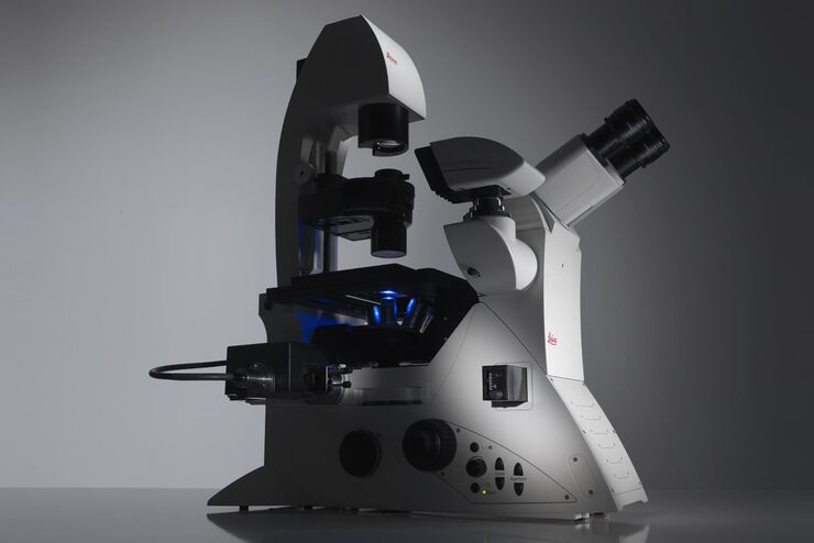
Leica DM4 B & DM6 B Upright Microscopes
Intelligent Automation for Life Science and Clinical Applications
Read our latest articles

Epi-Illumination Fluorescence and Reflection-Contrast Microscopy
This article discusses the development of epi-illumination and reflection contrast for fluorescence microscopy concerning life-science applications. Much was done by the Ploem research group…

Differential Interference Contrast (DIC) Microscopy
This article demonstrates how differential interference contrast (DIC) can be actually better than brightfield illumination when using microscopy to image unstained biological specimens.
 cells taken with phase contrast.")
Phase Contrast and Microscopy
This article explains phase contrast, an optical microscopy technique, which reveals fine details of unstained, transparent specimens that are difficult to see with common brightfield illumination.
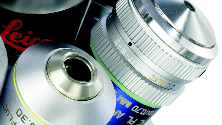
Immersion Objectives
How an immersion objective, which has a liquid medium between it and the specimen being observed, helps increase the numerical aperture and microscope resolution is explained in this article.

Hydraulics in the Automotive and Aerospace Industries
Cleanliness standards relating to lubricants, hydraulic fluids, and oils, e.g., ISO 4406 and DIN 51455 are discussed in this article. Cleanliness plays a central role in the automotive and…
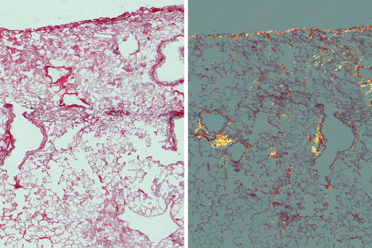
Studying Pulmonary Fibrosis
The results shown in this article demonstrate that fibrotic and non-fibrotic regions of collagen present in mouse lung tissue can be distinguished better with polarized light compared to brightfield.…
Factors to Consider When Selecting a Research Microscope
An optical microscope is often one of the central devices in a life-science research lab. It can be used for various applications which shed light on many scientific questions. Thereby the…
The Fundamentals and History of Fluorescence and Quantum Dots
At some point in your research and science career, you will no doubt come across fluorescence microscopy. This ubiquitous technique has transformed the way in which microscopists can image, tag and…

Koehler Illumination: A Brief History and a Practical Set Up in Five Easy Steps
In this article, we will look at the history of the technique of Koehler Illumination in addition to how to adjust the components in five easy steps.
 illuminated with wide-band UV excitation. Note the tissue structure with blue autofluorescence. Right: Same tissue and same immunostaining with FITC label illuminated with epi-illumination using narrow")
Milestones in Incident Light Fluorescence Microscopy
Since the middle of the last century, fluorescence microscopy developed into a bio scientific tool with one of the biggest impacts on our understanding of life. Watching cells and proteins with the…

Video Tutorial: How to Align the Bulb of a Fluorescence Lamp Housing
The traditional light source for fluorescence excitation is a fluorescence lamp housing with mercury burner. A prerequisite for achieving bright and homogeneous excitation is the correct centering and…

Video Tutorial: How to Change the Bulb of a Fluorescence Lamp Housing
When applying fluorescence microscopy in biological applications, a lamp housing with mercury burner is the most common light source. This video tutorial shows how to change the bulb of a traditional…

Fluorescent Proteins - From the Beginnings to the Nobel Prize
Fluorescent proteins are the fundament of recent fluorescence microscopy and its modern applications. Their discovery and consequent development was one of the most exciting innovations for life…

Optical Contrast Methods
Optical contrast methods give the potential to easily examine living and colorless specimens. Different microscopic techniques aim to change phase shifts caused by the interaction of light with the…

Polarization Contrast
Polarization microscopy is routinely applied in material sciences and geology to identify minerals on the basis of characteristic refraction properties and colors. In biology, polarization microscopy…

Fluorescence in Microscopy
Fluorescence microscopy is a special form of light microscopy. It uses the ability of fluorochromes to emit light after being excited with light of a certain wavelength. Proteins of interest can be…

Forensic Detection of Sperm from Sexual Assault Evidence
The impact of modern scientific methods on the analysis of crime scene evidence has dramatically changed many forensic sub-specialties. Arguably one of the most dramatic examples is the impact of…
Fields of Application
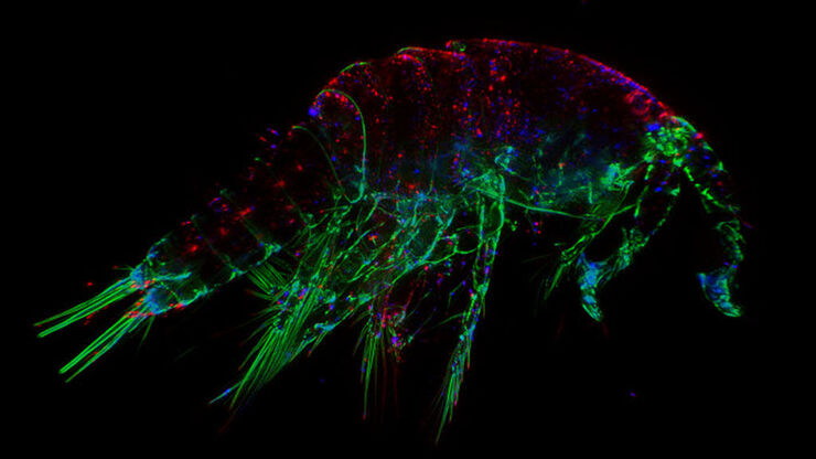
Fluorescence
Find out how fluorescence microscopes from Leica Microsystems support your research. Fluorescence is one of the most commonly used physical phenomena in biological and analytical microscopy for its…
Virology
Do your research interests focus on viral infection and disease? Find out how you can gain insights into virology with solutions for imaging and sample preparation from Leica Microsystems.
Basic Microscopy Techniques
Basic microscopy techniques are used in instances where the entire specimen on the microscope stage is exposed to a light source. The whole specimen is illuminated by white light either from above (in…
DIC Microscopes
A DIC microscope is a widefield microscopy which has a polarization filter and Wollaston prism between the light source and condenser lens and also between the objective lens and camera sensor or…
Darkfield Microscopes
The darkfield contrast method exploits diffraction or scattering of light from structures of a biological specimen or non-uniform features of a material sample.

Interested to know more?
Talk to our experts.
Do you prefer personal consulting? Show local contacts