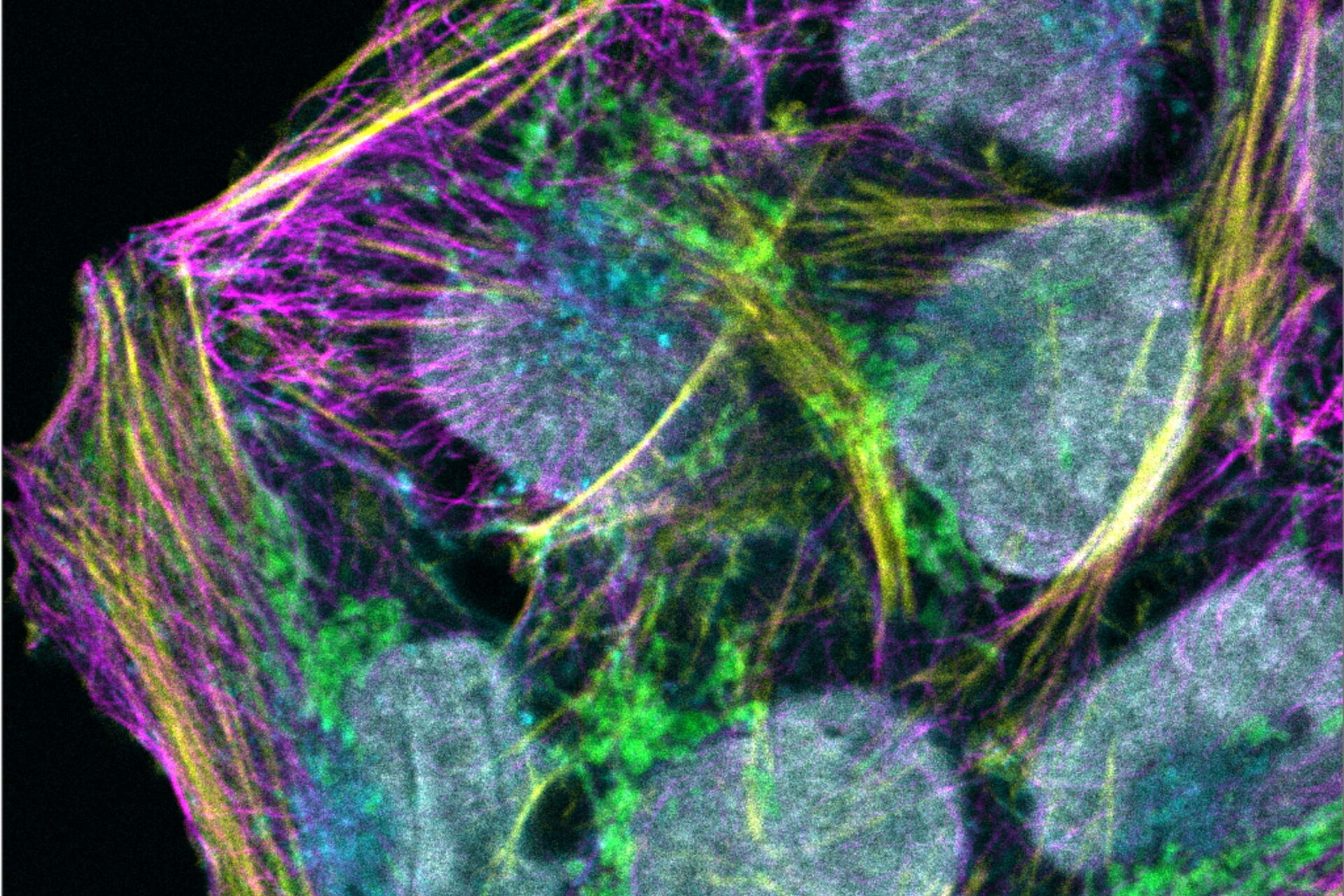

Content
Read the application note to find out how to:
- achieve complete wavelength freedom thanks to the unique combination of high-efficiency pulsed WLL excitation, AOBS technology, and spectral detection.
- get more flexibility when designing your experiment with an expanded WLL spectrum.
- use fluorescence-lifetime-derived information delivered by TauSense and increase the number of fluorescent probes which can be resolved in a single sample.
Download the PDF
Related Articles
-

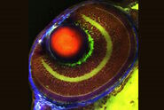
Extended Live-cell Imaging at Nanoscale Resolution
Extended live-cell imaging with TauSTED Xtend. Combined spatial and lifetime information allow…
Mar 05, 2024Read article -
 dataset, showing the biochemically distinct structures of a fresh, untreated apple slice.")
How to Prepare Samples for Stimulated Raman Scattering (SRS) imaging
Find here guidelines for how to prepare samples for stimulated Raman scattering (SRS), acquire…
Feb 05, 2024Read article -
Coherent Raman Scattering Microscopy Publication List
CRS (Coherent Raman Scattering) microscopy is an umbrella term for label-free methods that image…
Sep 11, 2023Read article
