Contact Us

Jennifer Kulhei , Dr. biol. hom.

Jennifer Kulhei

Jennifer Kulhei studied biochemistry, biophysical chemistry and animal physiology at the Goethe University in Frankfurt am Main. In the course of her diploma thesis, she focused on the development of a segmental isotopic labeled membrane protein for NMR spectroscopy. In her dissertation at the Max Planck Institute for Heart and Lung Research in Bad Nauheim she examined the dedifferentiation of adult mouse cardiomyocytes post myocardial ischemia utilizing transgenics, widefield and confocal microscopy as well as establishing a unique live-cell-sorting approach followed by next-generation sequencing to profile this rare cell type post myocardial infarction. In September 2018 she joined the Product Management Team at Leica Microsystems and is responsible for the inverted Widefield microscopy portfolio including the DMi8 based THUNDER Imagers.
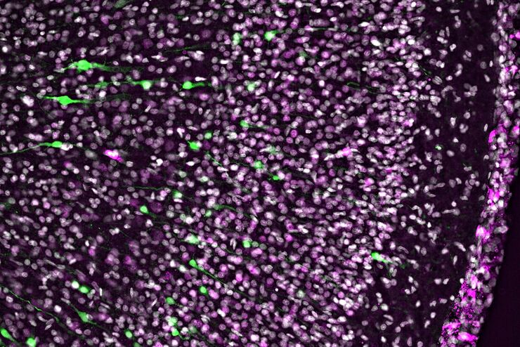
Electroporated nerve cells (green), specific neuronal markers (magenta) and cell nuclei (white), computational cleared.

Into the Third Dimension with "Wow Effect"- Observe Cells in 3D and Real-Time

Life is fast, especially for a cell. As a rule, cells should be examined under physiological conditions which are as close as possible to their natural environment. New technologies offer tremendous…
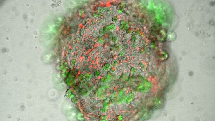
Lung organoid taken at the "liquid-air interface" with a THUNDER Imager 3D Cell Culture. The cells originate from transgenic mice, so that the different fluorescence represents the degree of differentiation of the respective cell (superposition). The image acquisition was performed on day 21 after the start of the culture. Reference: P. Kanrai, MPI-HLR Bad Nauheim.

Observing 3D Cell Cultures During Development

3D cell cultures, such as organoids and spheroids, give insights into cells and their interactions with their microenvironment. These 3D cell cultures are playing an increasingly important role for…
Scroll to top