Key Learnings
Dr. Tahiri and his team use the Leica EnFocus intraoperative OCT imaging system built into the Proveo 8 ophthalmic microscope, which provides greater insight during eye surgery. This cutting-edge technology allows surgeons to visualize subsurface tissue details, offering real-time intraoperative confirmation of the way tissue reacts during specific surgical maneuvers.
In the video presented here, Dr. Tahiri explains the benefits of using the EnFocus intraoperative OCT system for cataract incision analysis. He explains the advantages of using this imaging system, including accurate assessment of the incision architecture throughout the surgical procedure, which allows him to achieve optimal precision during cataract surgery.
You can find the video below, in addition to a full transcript in English. To learn more about the EnFocus intraoperative OCT or to find the right ophthalmic microscope for your needs, please contact a Leica representative. Our team will be happy to answer any of your questions and organize a personalized product demonstration.
About EnFocus
EnFocus is an intraoperative OCT for surgical microscopes. It allows to visualize subsurface details with high definition real-time images of the posterior and anterior segments. In addition to its benefits in keratoplasty and cataract surgery, intraoperative OCT can aid decision-making in cornea, glaucoma and retina procedures. It is also an essential tool in the application of gene therapies. EnFocus can be combined with the Leica Proveo 8 or M844 ophthalmology microscope for comprehensive visualization.
For more information about the Leica EnFocus intraoperative OCT or how to choose an ophthalmic microscope, contact a Leica representative.
Disclaimer: The statements and explanations of the healthcare professional in this video reflect only his opinion and personal experience. His statements don’t necessarily reflect the opinion of any institution with whom he is affiliated.
About Dr. Tahiri

Dr. Rachid Tahiri, a graduate of Paris VII University, France, is an ophthalmic surgeon working mostly in cataract surgery, retinal surgery, and clinical research. He is a Fellow of the European Board of Ophthalmology (FEBO) and holds a license from the Medical Council of Canada. He has contributed to several articles published in international peer-reviewed journals and has presented numerous scientific studies and posters at international ophthalmology meetings in Europe and the United States. Dr. Rachid TAHIRI is one of the co-organizers of Practical Ophthalmology Webinars, and one of the four artificial intelligence beta testers in France in diabetic retinopathy.
Transcription of the Presentation
Introduction
[00:06 - 00:26]
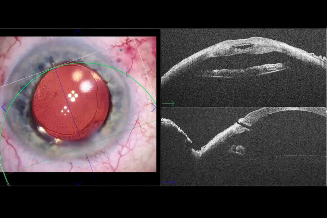
My name is Rachid Tahiri. I use intraoperative OCT Leica Deep EnFocus for one year. Here, I present another interesting use of intraoperative OCT. It might provide a valuable information about incision construction in cataract surgery.
Incision structure analysis from the start of surgery
[00:27 - 00:53]
In these examples, intraoperative OCT can give accurate and precise information about the incision architecture from the start of the surgery. We can clearly determine the incision length, the thickness of both superficial and deep flaps. We can perform analysis both in the longitudinal and transversal sections.
Incision analysis: before hydrosuture
[00:54 - 01:08]
Here, we can analyze the architecture of the incision at the end of the surgery. Before the hydrosuture, we note that the incision is discretely ovalized on the transversal section.
Incision analysis: during hydrosuture
[01:09 - 01:28]
In this example, the incision is more remodeled. We observe a more marked ovalization in the transversal section. In the longitudinal section, we observe a more direct incision with an internal and an external gap.
Incision analysis: during and after hydrosuture
[01:29 - 01:53]
We can use intraoperative OCT to analyze changes in the architecture of incision during hydrosuture. We note that the path of the incision disappears, embedded in the corneal edema. Here, we can see an incision before hydrosuture and after hydrosuture.
Incision analysis: characteristics of non-sealed incisions
[01:54 - 02:09]
When we analyze a sealed incision, the path of the incision is not visible. We note that the cornea’s anterior curvature remains regular, while a bulging accompanies the corneal edema of the posterior cornea.
[02:10 - 02:24]
For non-sealed incisions, despite the completion of the hydrosuture, we note in this case the presence of an internal gaping with a localized Descemet detachment.
[02:25 - 02:44]
In this case, despite the hydrosuture, the path of the incision remains visible. It is interesting to make a transversal section. In this case, we observe that the incision architecture is inclined. That explains why the incision was not sealed.
Incision analysis: after suturing
[02:45 - 03:08]
When suturing a non-sealed incision, its path becomes invisible. It is especially noted that the curvature of the anterior face remains almost unchanged. Then, the modification of curvature mainly concerns the posterior face, which becomes a double bump.
Conclusion: improving the quality of cataract surgery
[03:09 - 03:31]
In my experience, intraoperative OCT allows very interesting information about incision architecture. It is a great tool that allows finer and more precise analysis. This can improve the quality, the precision and the perfection of cataract surgery.
Related Articles
-
RPE65 Gene Therapy Subretinal Injection: Benefits of Intraoperative OCT
Discover how RPE65 gene therapy subretinal injection procedures in patients with Leber congenital…
Feb 01, 2024Read article -

Dislocated Cataract Angle Closure Aided by Intraoperative OCT
Learn how a dislocated cataract was treated with angle closure assisted by intraoperative OCT to…
Dec 07, 2023Read article -

Glaucoma Stent Revision Surgery Guided by Intraoperative OCT
Learn about a glaucoma subconjunctival stent revision guided by intraoperative OCT and the important…
Nov 28, 2023Read article
