Video Interview
Transcript of the video-interview
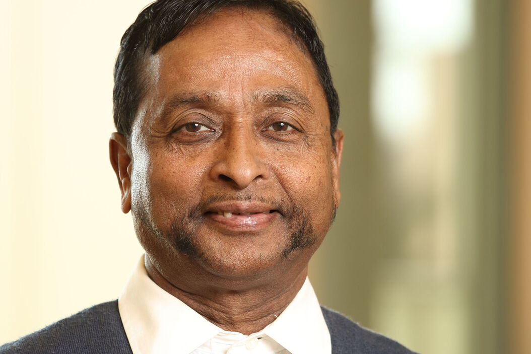
Dr. Ossato: Good afternoon Mr. Periasamy, thank you very much for being here and having agreed to this interview regarding functional and fluorescence lifetime imaging. Last month Alessandro Esposito (Medical Research Council, University of Cambridge, UK) posted the most prolific authors of lifetime imaging and you are the fifth best, so congratulations!
Prof. Periasamy: Thank you!
Dr. Ossato: And would you mind to share with us some of your research in functional imaging?
Prof. Periasamy: Thank you very much for inviting me! It is always a great pleasure to be here to talk about the lifetime for functional imaging. I have done a lot of work on the research side of using the lifetime. Currently I am working on fluorescence lifetime redox ratio using endogenous molecules to measure the metabolism and also mitochondrial disfunction in live cells, tissues, and animals. That’s my main research right now I am working on.
Dr. Ossato: So everything label free. So you are able to follow metabolism without any labeling or extra standard fluorescence dye?
Prof. Periasamy: That is correct. It is that NADH, FAD, and tryptophan provide autofluorescence. You do not have to label it. So this kind of technique is useful for translational research. More importantly, lifetime is the technique that possibly you can apply for this kind of measurements because it provides info for the bound and free coenzymes NADH and FAD. So it is only possible to discriminate these two functions bound and free using the lifetime technique. With the intensity technique it is impossible to do that.
Dr. Ossato: Okay, thank you for the explanation. What do you think were the most relevant developments in lifetime imaging in the last 15 years?
Prof. Periasamy: Well, so the lifetime imaging more or less started in the 1990s. Before that, the lifetime technique was available only in chemistry and physics labs. The important thing we have to think about is the interpretation of lifetime for biological data. That is where the biologists are basically looking for how I can connect the lifetime measurements to the biology. So that requires some kind of education to educate them how the lifetime technique is maybe useful for biology.
Dr. Ossato: So helping also in the interpretation of the data, where the molecular interaction that changes the lifetime, for example, is coming from.
Prof. Periasamy: That’s correct. The lifetime technique is very well used in the case of molecular interaction, protein-protein interaction, which is called a FRAP. So that is called a lifetime technique which is advanced.
Dr. Ossato: So there were a lot of developments in the last years. What are we still missing for the fluorescence lifetime imaging part?
Prof. Periasamy: So, the still missing part in the lifetime measurement is the speed and also the data analysis, because the biology is the nanosecond event. We can measure the lifetime in the nanosecond time mode. But the technology has to advance to speed up the data collection and also not to photobleach. And in real time, if we display the data analysis board while collecting the lifetime, the biologists can easily interpret the results of the measurements.
Dr. Ossato: So you think lifetime has still a lot of place in the biology lab and helping with a new technology will spread this new contrast fluorescence microscopy.
Prof. Periasamy: That’s correct. The lifetime imaging is a very good contrast technique. And not only contrast, also you can do the dynamic meaning even when the protein-protein molecules interact. So this is a dynamic technique and it will advance pretty sure. It will be available in all biology labs and microscopy facilies like a confocal. I think, in my opinion, the FALCON system satisfies that condition. But still, you know, the biologists will have to catch up with what’s going on.
Dr. Ossato: We need to educate them. We should have some workshops soon. Thank you very much for your time it was really a pleasure talking to you today.
Prof. Periasamy: Thank you very much for your time.
Related Articles
-

Live-Cell Fluorescence Lifetime Multiplexing Using Organic Fluorophores
On-demand video: Imaging more subcellular targets by using fluorescence lifetime multiplexing…
Nov 18, 2022Read article -

What is FRET with FLIM (FLIM-FRET)?
This article explains the FLIM-FRET method which combines resonance energy transfer and fluorescence…
Nov 15, 2022Read article -

Visualizing Protein-Protein Interactions by Non-Fitting and Easy FRET-FLIM Approaches
The Webinar with Dr. Sergi Padilla-Parra is about visualizing protein-protein interaction. He gives…
Nov 13, 2022Read article

