Related Articles
-

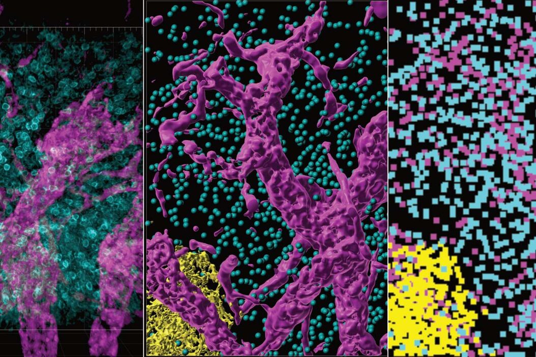
IBEX, Cell DIVE, and RNA-Seq: A Multi-omics Approach to Follicular Lymphoma
In a recent study by Radtke et al., a multi-omics spatial biology approach helps shed light on early…
Apr 08, 2024Read article -

Notable AI-based Solutions for Phenotypic Drug Screening
Learn about notable optical microscope solutions for phenotypic drug screening using 3D-cell…
Dec 06, 2023Read article -

Understanding Tumor Heterogeneity with Protein Marker Imaging
Explore tumor heterogeneity and immune cell dynamics. See how quantitative imaging analysis reveals…
Dec 04, 2023Read article
Related Pages
-
THUNDER Imagers: High Performance, Versatility and Ease-of-Use for your Everyday Imaging Workflows
This webinar will showcase the versatility and performance of THUNDER Imagers in many different life…
Visit related page -

Computational Clearing - Enhance 3D Specimen Imaging
This webinar is designed to clarify crucial specifications that contribute to THUNDER Imagers'…
Visit related page
