Contact Us

How to Successfully Implement Coral Life

 How_to_successfully_implement_the_Leica_Nano_workflow_teaser.jpg

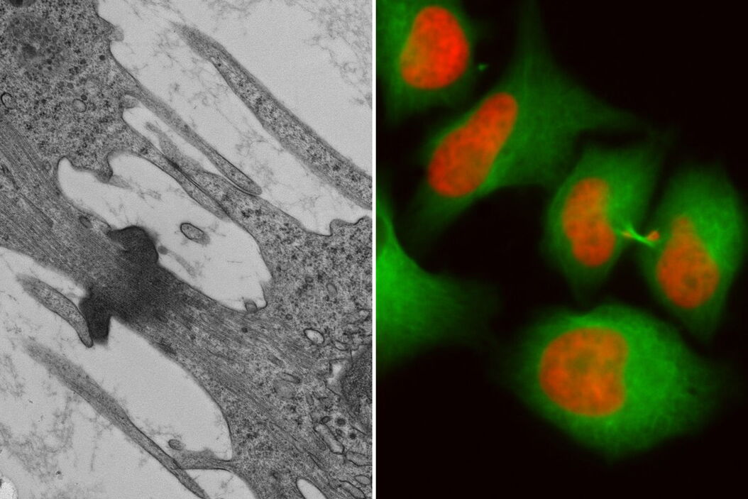
Many electron microscope (EM) workflows start with sample fixation followed by sample preparation and EM imaging. However, samples displaying interesting behavior tend to be rare and finding the “right cell” can be time-consuming and tedious. The live-cell  CLEM workflow allows you to capture dynamic information related to a relevant biological process as it happens and put these observations into their ultrastructural context. The Coral Life workflow streamlines this process to optimize your performance and increase your productivity. In this webinar, we will demonstrate the Coral Life workflow using an example which shows how reproducible results can be obtained without compromising sample quality.

Please note that the Leica Nano Workflow was renamed to Coral Life. The previous name Leica Nano Workflow is still used within the recorded webinar. 

Key Learnings

  • How to maintain physiological conditions from cell culture until cryo fixation
  • How to optimize your live-cell imaging for live-cell CLEM applications
  • Easy transfer from the THUNDER Imager Nano to the EM ICE high pressure freezer
  • Retrieve your cell of interest in the resin block

Register to watch the webinar

By clicking on the SUBMIT button, I confirm that I have reviewed and agree with Leica Microsystems GmbH Terms of Use and their Privacy Policy. I also understand my privacy choices as they pertain to my personal data, as detailed in the aforementioned Privacy Policy under ‘’Your Privacy Choices’’.

Related Articles

Background image
Scroll to top