
The difficulties can be overcome with a combination of fluorescence light sheet microscopy technologies and optical clearing methods. In a recent study, several metrics for evaluating the efficiency of clearing protocols are compared. An imaging guideline for reaching single-cell resolution when analyzing multicellular aggregates with the SP8 DLS microscope system is also described.
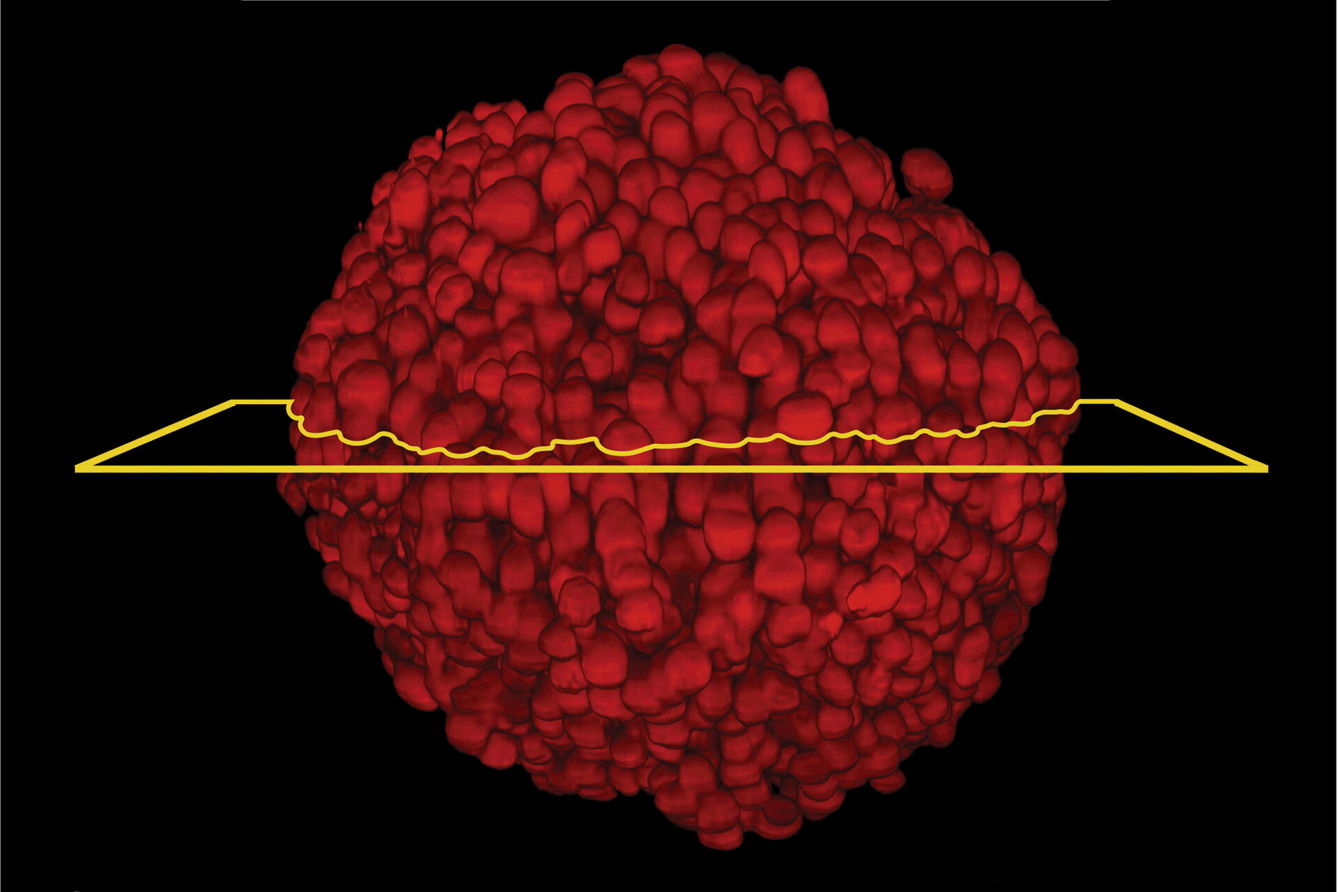
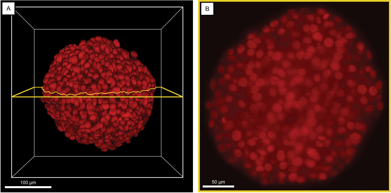
One of the DLS main advantages is that it facilitates illumination of the sample from two sides. Spheroids with a diameter of around 250 µm, derived from 3 different cell lines, were analyzed and details inside them visualized using 5 different clearing protocols. The full 3D dataset, which includes 90 fluorescently stained spheroids, has been shared with the community. To characterize the image quality of spheroids, organoids, or other 3D multicellular aggregates, a novel metric is proposed. This metric can serve as a gold standard for the quantitative comparison of optical clearing protocols and help users in choosing the most suitable one for any given experiment.
A Diosdi, D Hirling, M Kovacs, T Troth, M Harmati, K Koos, K Buzas, F Piccinini, P Horvath:
A quantitative metric for the comparative evaluation of optical clearing protocols for 3D multicellular spheroids
Computational and Structural Biotechnology Journal, 19:1233-1243, February 2021. DOI: 10.1016/j.csbj.2021.01.040
https://www.sciencedirect.com/science/article/pii/S2001037021000441
Related Articles
-

Extended Live-cell Imaging at Nanoscale Resolution
Extended live-cell imaging with TauSTED Xtend. Combined spatial and lifetime information allow…
Mar 05, 2024Read article -
 dataset, showing the biochemically distinct structures of a fresh, untreated apple slice.")
How to Prepare Samples for Stimulated Raman Scattering (SRS) imaging
Find here guidelines for how to prepare samples for stimulated Raman scattering (SRS), acquire…
Feb 05, 2024Read article -
Coherent Raman Scattering Microscopy Publication List
CRS (Coherent Raman Scattering) microscopy is an umbrella term for label-free methods that image…
Sep 11, 2023Read article
