Stem Cells
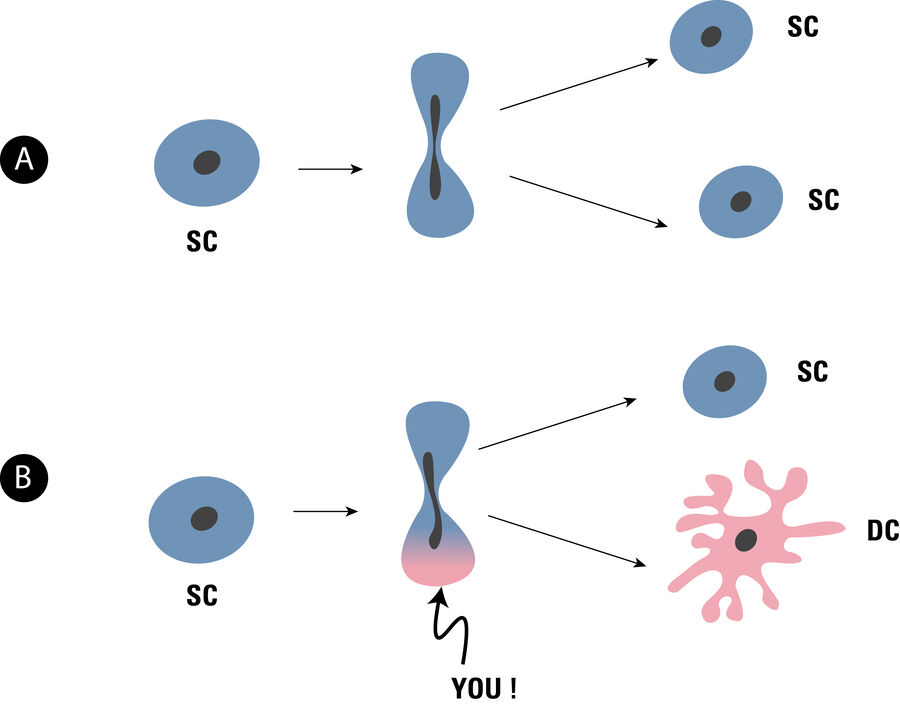
In order to fulfill these functions, the stem cells divide either symmetrically (and thus supply new stem cells), or asymmetrically, creating a stem cell and a cell that differentiates in further divisions to the desired tissue cell. Originally it was assumed that for the proliferation of tissue cells necessarily an asymmetric division is necessary, but it is now known that there are also alternative cell division concepts in which only symmetric cell divisions occur.
Depending on which differentiation options a stem cell still holds, a distinction is made between different stages: Totipotent stem cells can be used to create a new organism. Pluripotent stem cells can deliver different tissue cells. Multipotent stem cells deliver different cell types, but only for a specific tissue.
Niches
The above subdivision has now been called into question because it has been found that multipotent stem cells are also able to deliver cells for quite different tissues when implanted there. One explanation probably provides the insight that the stem cells are not freely present in the tissue, but are associated with specific cells: the stem cells are located in a microenvironment, called “niche”, quite comparable to a wall niche. This niche ensures that, for example, asymmetric division is possible. The niche can also determine the direction of differentiation.
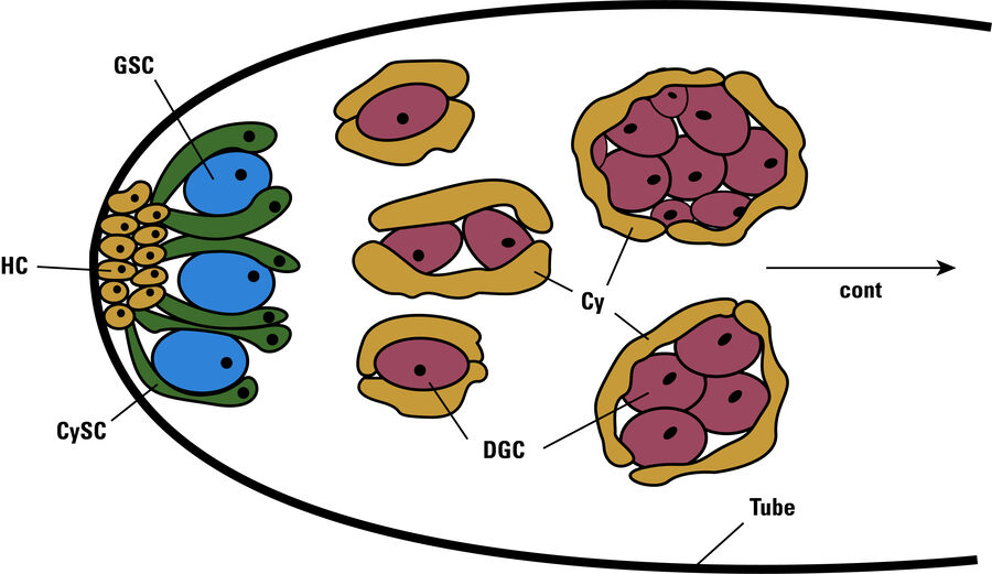
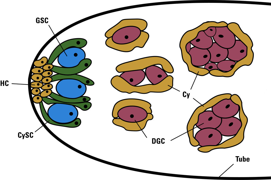
The testicles of the male fruit flies (Drosophila melanogaster) are elongated tubes which are rolled up helically. The niche for the reproductive cells is located at the front end of these tubes. At the very end of the tube, there are support cells (hub cells), which support two different stem cells. One, of course, the stem cells of the germ line, from which sperm cells are to be derived, on the other hand, cyst stem cells, from which later develop cysts, in which germinate the cells, see Figure 2.
Niche-Microscopy
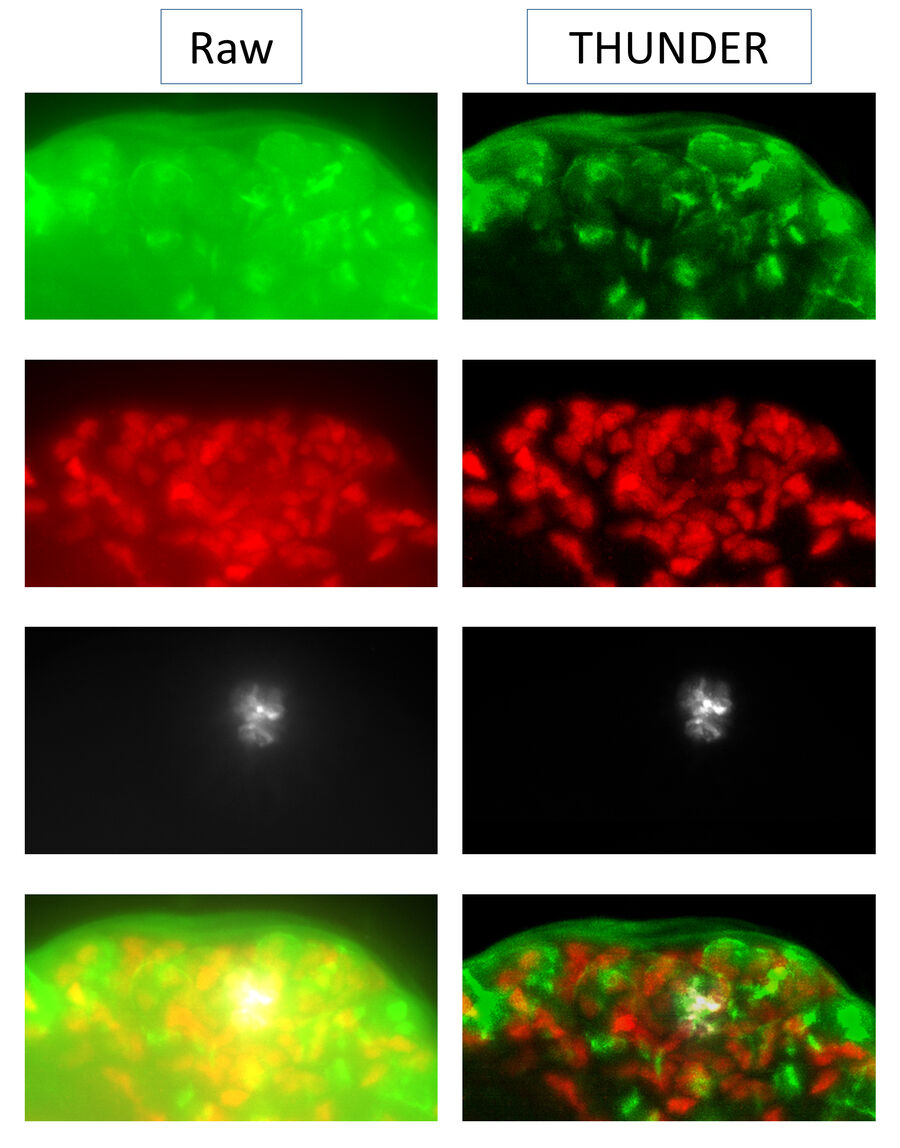
The research goal of Dr. Kari Lenhart at Drexel University Biology Department is to understand further how stem cells control normal tissue renewal. Dr. Lenhart performs her studies using the drosophila testis niche. Three channel x,y,z stacks were collected from drosophila testis at the stem cell niche and includes the two stem cell populations; the somatic cyst stem cells and the germline stem cells.
The DMi8 microscope from Leica Microsystems with a 63x/1.2 water objective, DFC9000 sCMOS camera and cool LED fluorescence illuminator was used to collect the data set. Superior image quality is unparalleled in widefield microscopy due to the high quality of the optics throughout the system.
The THUNDER Computational Clearing shows details in the three-dimensional sample, that are not available by classical fluorescence microscopy. For a comparison see Figure 3.
Related Articles
-

Overcoming Observational Challenges in Organoid 3D Cell Culture
Learn how to overcome challenges in observing organoid growth. Read this article and discover new…
Apr 08, 2024Read article -

How do Cells Talk to Each Other During Neurodevelopment?
Professor Silvia Capello presents her group’s research on cellular crosstalk in neurodevelopmental…
Apr 03, 2024Read article -

Technical Terms for Digital Microscope Cameras and Image Analysis
Learn more about the basic principles behind digital microscope camera technologies, how digital…
Dec 14, 2023Read article
