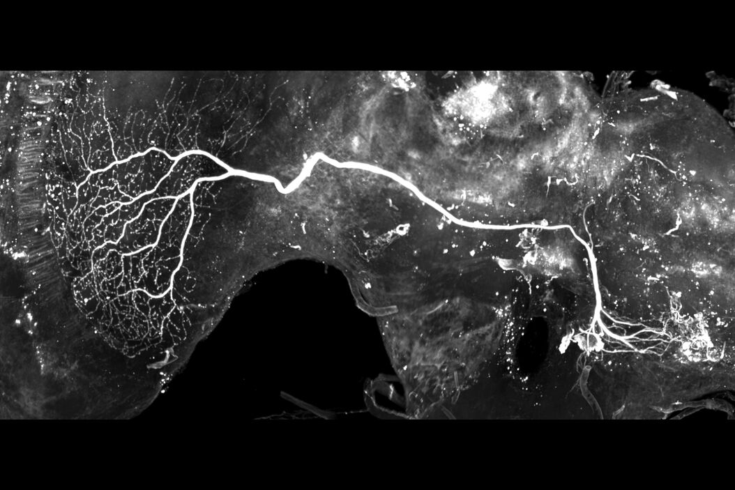
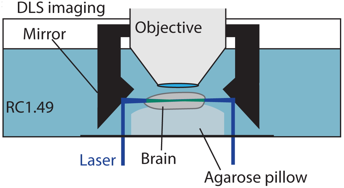
Brains were imaged with the Digital LightSheet (DLS) from Leica Microsystems. It allows the user to choose between several detection objectives and mounting frames enabling high-resolution visualization. Users can also take full advantage of all the structural details revealed within the 3D cleared insect brains.
Bekkouche B.M.B., Fritz H.K.M., Rigosi E. & O'Carroll D.C.:
Comparison of Transparency and Shrinkage During Clearing of Insect Brains Using Media With Tunable Refractive Index
Front. Neuroanat., 20 November 2020, https://doi.org/10.3389/fnana.2020.599282
https://www.frontiersin.org/articles/10.3389/fnana.2020.599282/full
Related Articles
-

Overcoming Observational Challenges in Organoid 3D Cell Culture
Learn how to overcome challenges in observing organoid growth. Read this article and discover new…
Apr 08, 2024Read article -
 imaged with the THUNDER Imager 3D Cell Culture. Courtesy of Dr. F.T. Arroso Martins, Tamere University, Finland.")
How to Get Deeper Insights into your Organoid and Spheroid Models
In this eBook, learn about key considerations for imaging 3D cultures, such as organoids and…
Nov 22, 2023Read article -
 acquired using THUNDER Imager Live Cell. Image courtesy of Janina Kaspar and Irene Santisteban, Schäfer Lab, TUM.")
Imaging Organoid Models to Investigate Brain Health
Imaging human brain organoid models to study the phenotypes of specialized brain cells called…
Jul 11, 2023Read article
