
The challenges with out-of-focus blur and long acquisition times
In conventional widefield imaging, a common issue is out-of-focus blur, especially when imaging thick sections or whole-mounted samples like the retina. Blur is caused by illumination of the whole sample, particularly the areas above and below the image plane. This causes poor contrast of fluorophore channels, masks image information, and reduces the ability for clear and reproducible analysis. Confocal point scanners allow optical sectioning and are the benchmark for superior image quality and high-contrast fluorescence images, even for multicolour data acquisition. However, with confocal point scanners, acquiring image sequences of large samples might be time consuming depending on the scanning speed.
Together, low-throughput acquisition and blur can hamper a scientist’s ability to quantify cellular and molecular features of tissue like the retina. For this type of research, it’s important to have a solution capable of capturing images with high acuity and fast screening of the retina, so 3D reconstruction can be performed.
High-acuity imaging of whole-mounted retina samples using THUNDER
In previous experiments using widefield imaging, 300-micron thick sections were imaged as mosaics at 10x magnification, on a single plane, and were generated from >100 tiles to create a composite image. The aim was to quantify cell damage caused by retinal degeneration. Two model animal species were used for the experiments: Control Swiss adult female mouse and Control Sprague Dawley adult female rats.
First, the retinas were dissected, whole-mounted and processed for immunohistochemistry experiments. Antibodies against two different proteins were used to assess cell damage: Iba1, a marker for microglial cells (labelled with Alexa Fluor® 488 green-fluorochrome), and Brn3a to target retinal ganglion cells (labelled with Alexa Fluor® 594 red-fluorochrome). Samples were then imaged with THUNDER at 20x for mice retina and 10x for rat retina.
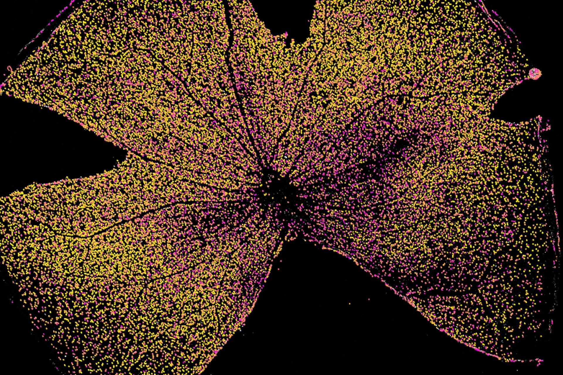
Imaging with THUNDER allowed rapid, high contrast images of the whole-mounted retina. In contrast, previous experiments using widefield imaging created results with considerable amounts of blur, and poor contrast of red and green channels (Figure 1A). When imaged with THUNDER, combined with its Instant Computational Clearing (ICC), the quality of the image is much improved (Figure 1B). There is far less blur, the contrast is higher, and there is a much clearer snapshot of the cellular integrity of the retina.
 and THUNDER (B) snapshot of the whole-mounted retina. Microglia (Iba1, green) and retinal ganglion cells (Brn3a, red). Sample courtesy of the Experimental Ophthalmology Group, University of Murcia, Spain.")
Powerful image analysis of THUNDERed images with Aivia
In the next section we show how AI based analysis combined with Computationally Cleared Images (THUNDERed images) produces high confidence results. The images of the retina captured with THUNDER can be analyzed with Aivia without the need of any post acquisition processing steps, saving time and effort in the imaging workflow, while increasing accuracy of analysis. Acquiring the whole-mounted retina on a THUNDER imager reduces background blur, resulting in higher clarity images that allow for accurate segmentation with Aivia. In this experiment, it was possible to produce reliable reconstructions of highly complex retina samples with the combination of THUNDER & Aivia.
As an example, the raw image in Figure 2A has high, variable background signal, with corresponding signal peak intensities increasing with background intensity (Figure 2B). After Computational Clearing using THUNDER, the background is removed (Figure 2C) and signal peak intensities can now easily be quantified and compared (Figure 2D). This allows for more accurate analysis with Aivia later in the workflow.
 Raw image with high inhomogeneous background and (B) corresponding signal peak intensities. (C) In the THUNDERed image the background is removed. The signal peak intensities (D) can easily be quantified and compared.")
The signal peak intensities (D) can easily be quantified and compared. Sample courtesy of the Experimental Ophthalmology Group, University of Murcia, Spain.
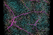
A detail view on the THUNDERed retina shows that background was effectively removed (Figure 3). The corresponding analysis shows the high quality of the Aivia object detection, performed with Aivia Machine Learning Pixel Classifier, on the THUNDERed image.
The Pixel Classifier enabled a classification confidence value to be calculated for each pixel, creating an average confidence per object detected. This data can then be plotted, to assess the overall quality of detection. Images analyzed with the Pixel Classifier can also be reported and visualized on top of the sample images, with a colour code demonstrating the confidence level of each classified object (Figure 4).

Confidence levels of the detected objects were subsequently plotted in a violin plot (Figure 5). The data shows that THUNDERed images (right) had significantly more objects at a higher confidence level compared to the violin plot of the raw image analysis (left).
The superior results generated in this experiment illustrated the use of THUNDER and Aivia to enhance the detection and precise quantification of individual cells. The creation of annotations to train Machine Learning in Aivia was able to be performed without any contrast adjustment, generating results in a simpler and faster way.

Conclusions
Results show that the THUNDER imaging platform is able to achieve fast and sharp imaging of whole-mounted thick sections of mouse retina. These improved imaging results, when compared to widefield microscopy, help to enable better quality and reproducible analysis of thick tissues like the retina.
Related Articles
-

Create New Options for Live Cell Imaging
The use of state-of-the-art AI systems is pushing image analysis into a new generation. Challenges…
Apr 08, 2022Read article -

Accurately Analyze Fluorescent Widefield Images
The specificity of fluorescence microscopy allows researchers to accurately observe and analyze…
Jan 17, 2022Read article -
Save Time and Effort with AI-assisted Fluorescence Image Analysis
The powerful synergy of THUNDER and Aivia analyze fluorescence images with greater accuracy, even…
Nov 16, 2021Read article
Related Pages
-
Microscope Imaging Software
Microscope imaging software from Leica Microsystems combines microscope, digital camera and…
Visit related page -
THUNDER Imaging Systems
To answer important scientific questions, THUNDER Imaging Systems enable you to obtain a clear view…
Visit related page
