
Now, STELLARIS Digital LightSheet combines a confocal system and a light sheet microscope into one instrument, giving you the flexibility you need to make groundbreaking discoveries and advance your research. With the TwinFlect mirrors designed by Leica Microsystems, the platform integrates light sheet’s power into a confocal platform. As well as 3D imaging of live biological samples, DLS is suitable for the efficient acquisition of cleared specimens – all in one system. This article explains how light sheet technology works, and how combining it with confocal microscopy can revolutionize your research and allow you to discover more.
Light sheet imaging: the basics
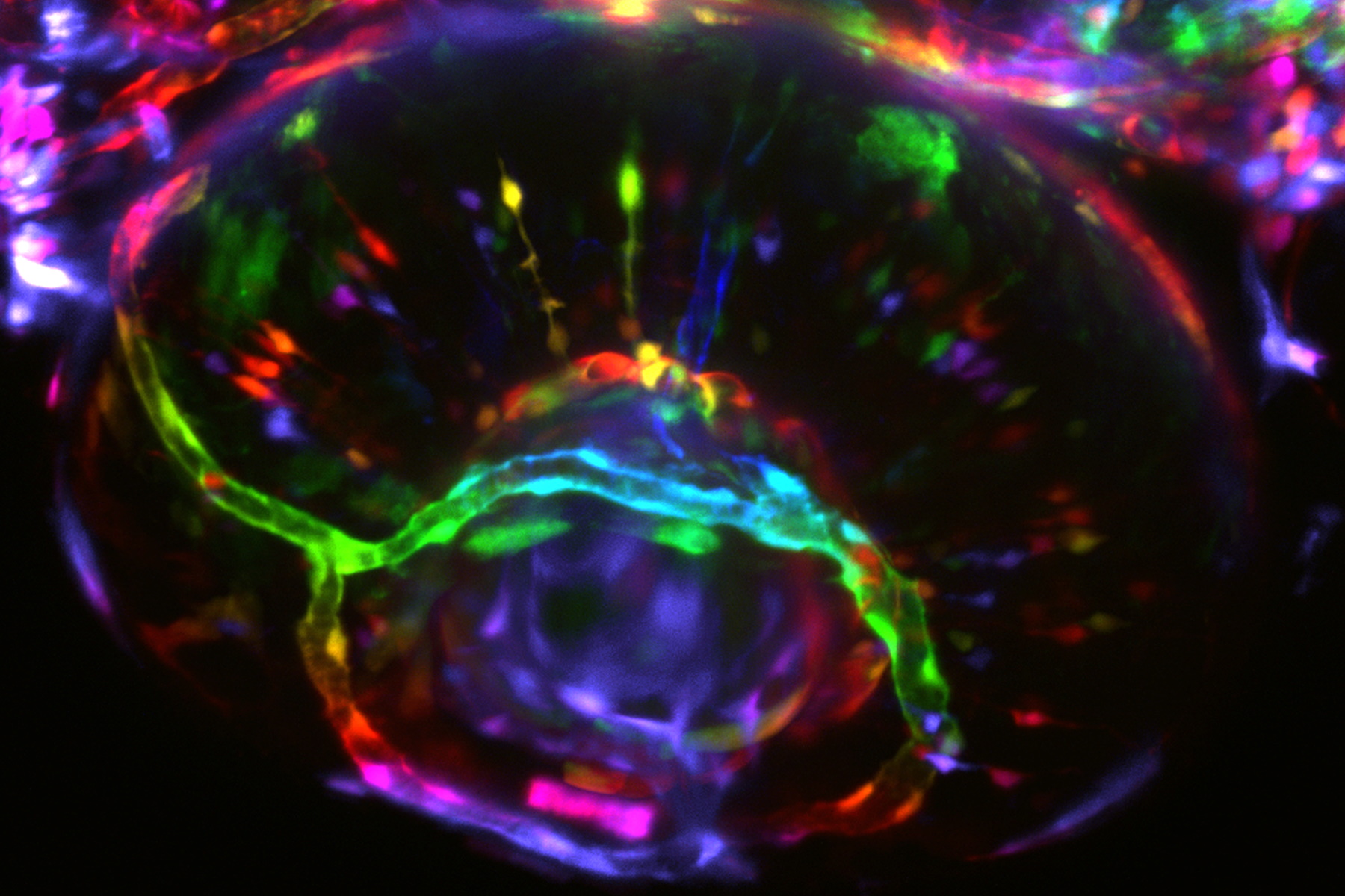
Working with biological samples can be complex, and optical imaging methods such as light sheet microscopy allow you to see the smaller details. Whether you’re following developing organisms over hours or even days, light sheet technology enables you to learn more about your specimen without any noticeable fluorescence signal weakening. But what makes light sheet microscopy so effective?
The power lies in the illumination and detection. Light sheet microscopes illuminate your specimen in a single plane, enabling you to image highly sensitive samples or fast biological processes. Your live specimens are imaged in 3D, as you automatically have optical sectioning when moving your sample through the light sheet. The plane is captured immediately using a camera – and it’s this parallelization of acquisition which allows rapid visualization of live sample dynamics.
Alongside sensitivity and speed, light sheet microscopy treats your samples with the care they require thanks to the restriction of light to one plane. Light sheet microscopy automatically increases your sample protection: there is no out-of-focus excitation, which reduces phototoxic effects to the focal plane. This lowers the overall photodamage to your sample from toxicity and bleaching.
In confocal imaging, the illumination path is shared with the detection path on the same axis [1], while in light sheet microscopy the illumination and detection objectives are perpendicular to each other. Both systems are powerful: confocal microscopy provides you with effortless analysis of your specimens in high resolution, while light sheet microscopy facilitates live imaging of thicker samples. Now, by virtue of technological advances, both methods can be combined into a single instrument with incredible results.
Microscopy options to transform your research
STELLARIS Digital LightSheet (DLS) is a platform that allows you to harness the benefits of both light sheet and confocal microscopy effectively. You can exploit the advantages of light sheet microscopy’s enhancement of your sample, and simultaneously retain confocal functionality. This is achieved by the unique TwinFlect mirror device – but how does it work?
The TwinFlect mirror device deflects the illuminating light sheet at a 90° angle, turning your device into a light sheet microscope. By this process, the TwinFlect mirrors grant the integration of the illumination and detection beam path into the vertical axis of every STELLARIS system with an inverted microscope stand. Moreover, both objectives and TwinFlect mirrors are easily exchanged, opening the path to reaching your goals more easily.
There’s more to STELLARIS DLS than meets the eye. As well as easy transition between your experiments, we explore further benefits that incorporating light sheet technology can bring.
Specialized light sheet optics
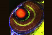
The objectives and the TwinFlect mirror device form the core of the DLS system. Superior objectives can be utilized for a wide range of applications, allowing you to explore different dimensions in one system. The system even grants visualization of structural details in cleared tissues and organisms, so you can find the information you need.
The STELLARIS DLS system enables flexibility in your research while providing you with choice. By choosing different illumination objectives optimized to the application need, you can shape the light sheet to meet your experimental requirements. Whether exploring the subtleties of your specimen, increasing your field of view, or adapting to the refractive index of your imaging medium, you can optimize your experiment in the DLS system to enhance the potential of your research. There is an expanding number of options available for your optimal detection objective, as shown below, serving application requirements from live samples to fixed and cleared specimens.
Objective | Live cell imaging | Sugar based clearing reagents | BAAB clearing compatible |
| FLUOTAR L 25x/0.95 W | Yes | No | No |
| APO L 20x/0.5 W | Yes | No | No |
| FLUOTAR L 16x/0.6 IMM | Yes | Yes | Yes |
| APO L 10x/0.3 | Yes | Yes | No |
| 5x/0.15 IMM | Yes | Yes | Yes |
Table 1: Objectives for light sheet microscopy with STELLARIS DLS.
Simple sample preparation
STELLARIS DLS is exceptionally easy to use: the vertical experimental set-up means you can keep your familiar sample preparation approach. Specimens can, for example, be embedded in agarose on standard glass-bottom petri dishes, leaving them directly accessible. The only consideration in this preparation? Remove excessive agar to make room for the TwinFlect mirror, and your experiment is ready to run.

The effortless configuration allows you to screen several samples in multi-positioning experiments. This is facilitated by the stage automation, enabling you to image several specimens in a single experimental set-up. Thanks to the DLS system, you can discover more in less time.
STELLARIS DLS facilitates sample mounting as well [2]. Glass capillaries act as an excellent scaffold for samples, allowing a lower percentage matrix to be used. There are two sizes of U-shaped glass capillaries available, meaning they can fit with different TwinFlect mirrors to give you perfect sample positioning. To help with positioning, a rotation device adjusts the viewing angle to give superior sample alignment, allowing users to choose which side of the sample to investigate [3]. FEP tubes, having a refractive index of 1,338, enable imaging in an aqueous environment.
You are also able to achieve more efficient DLS imaging by employing Leica mounting frames [4]. The frames enable preparation of more specimens and can be used with potentially harmful reagents such as BABB (benzyl alcohol benzyl benzoate), expanding your opportunity for experimental investigation.

Accomplish more with STELLARIS DLS
Light sheet microscopy is a mighty tool, whether you’re imaging sensitive specimens or fast dynamic processes. When integrated into a confocal system with TwinFlect mirrors, it provides researchers with the potential to find more answers. From live samples to fixed and cleared specimens, STELLARIS DLS provides you with a flexible solution for gentle 3D imaging of biological samples, with convenient sample mounting and superior optics.
References
- R. T. Borlinghaus, P. Haas, Confocal and Digital Light Sheet Imaging, Science Lab (2015) Leica Microsystems.
- P. Haas, DLS Sample Preparation: Using U-Shaped Glass Capillaries for Sample Mounting, Science Lab (2017) Leica Microsystems.
- P. Haas, E. R. Motta, L. O. Lopez, B. C. Velluttini, P. Tomancak, E. Reynaud, Using a Rotation Device for DLS Sample Mounting, Science Lab (2019) Leica Microsystems.
- S. Diehl, P. Haas, Using Mounting Frames for DLS Sample Preparation, Science Lab (2019) Leica Microsystems.
Related Articles
-

Extended Live-cell Imaging at Nanoscale Resolution
Extended live-cell imaging with TauSTED Xtend. Combined spatial and lifetime information allow…
Mar 05, 2024Read article -
 dataset, showing the biochemically distinct structures of a fresh, untreated apple slice.")
How to Prepare Samples for Stimulated Raman Scattering (SRS) imaging
Find here guidelines for how to prepare samples for stimulated Raman scattering (SRS), acquire…
Feb 05, 2024Read article -
Coherent Raman Scattering Microscopy Publication List
CRS (Coherent Raman Scattering) microscopy is an umbrella term for label-free methods that image…
Sep 11, 2023Read article
