Overview of the case study
- Initial patient presentation
- Pre-operative assessment
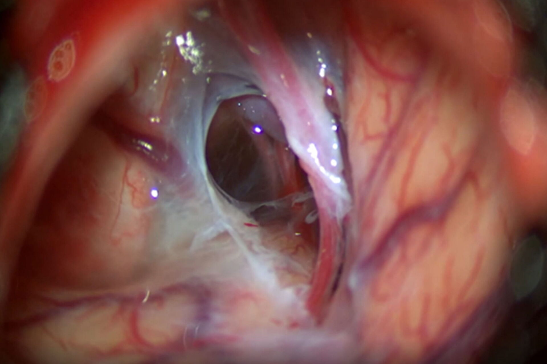
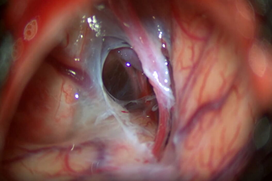
- Treatment decision and surgical procedure
- Post-operative assessment and imaging
- Impact of the M530 OHX surgical microscope on the treatment of the Galassi type III arachnoid cyst
Related Articles
-

How do Cells Talk to Each Other During Neurodevelopment?
Professor Silvia Capello presents her group’s research on cellular crosstalk in neurodevelopmental…
Apr 03, 2024Read article -

The Shape of the Brain: Spatial Biology of Alzheimer’s Disease
Uncover cell identity and brain structure in Alzheimer's disease with Cell DIVE multiplexed imaging,…
Nov 29, 2023Read article -
Coherent Raman Scattering Microscopy Publication List
CRS (Coherent Raman Scattering) microscopy is an umbrella term for label-free methods that image…
Sep 11, 2023Read article
