Key Learnings
The Proveo 8 ophthalmic microscope with built-in intraoperative OCT is an advanced imaging solution designed to improve the surgical workflow and achieve the highest level of precision during anterior and posterior segment surgery.
In the video below, Dr. Tahiri shares his impressions of using the Proveo 8 intraoperative OCT imaging system in cataract surgery. Specifically, Dr. Tahiri highlights the advantage of using this imaging solution in detecting and addressing residual lens fragments, which can be difficult to visualize due to being hidden behind surgical incisions or edema. He explains that using Proveo 8 intraoperative OCT is useful in locating residual lens fragments, improving surgical outcomes, and reducing the risk of postoperative complications and readmission for secondary surgery.
Discover the video below, as well as its transcript in English. To learn more about the Proveo 8 intraoperative OCT imaging system or to find the right ophthalmic microscope for your requirements, please contact a Leica representative. Our team will be happy to answer your questions and organize a personalized product demonstration for you.
About EnFocus
EnFocus is an intraoperative OCT for surgical microscopes. It allows to visualize subsurface details with high definition real-time images of the posterior and anterior segments. In addition to its benefits in keratoplasty and cataract surgery, intraoperative OCT can aid decision-making in cornea, glaucoma and retina procedures. It is also an essential tool in the application of gene therapies. EnFocus can be combined with the Leica Proveo 8 or M844 ophthalmology microscope for comprehensive visualization.
For more information about the Leica EnFocus intraoperative OCT or how to choose an ophthalmic microscope, contact a Leica representative.
Disclaimer: The statements and explanations of the healthcare professional in this video reflect only his opinion and personal experience. His statements don’t necessarily reflect the opinion of any institution with whom he is affiliated.
About Dr. Tahiri

Dr. Rachid Tahiri, a graduate of Paris VII University, France, is an ophthalmic surgeon working mostly in cataract surgery, retinal surgery, and clinical research. He is a Fellow of the European Board of Ophthalmology (FEBO) and holds a license from the Medical Council of Canada. He has contributed to several articles published in international peer-reviewed journals and has presented numerous scientific studies and posters at international ophthalmology meetings in Europe and the United States. Dr. Rachid TAHIRI is one of the co-organizers of Practical Ophthalmology Webinars, and one of the four artificial intelligence beta testers in France in diabetic retinopathy.
Transcription of the Presentation
Introduction
[00:06 - 00:17]
Hi, my name is Rachid Tahiri. I am an ophthalmic surgeon from Granville in Normandy, France. My main topics are cataract and retinal surgery.
Residual lens fragments assessment
[00:17 - 00:32]
Beyond retinal surgery, which is a classical indication, intraoperative OCT may be useful in cataract surgery, especially to access residual lens fragments.
[00:33 - 00:56]
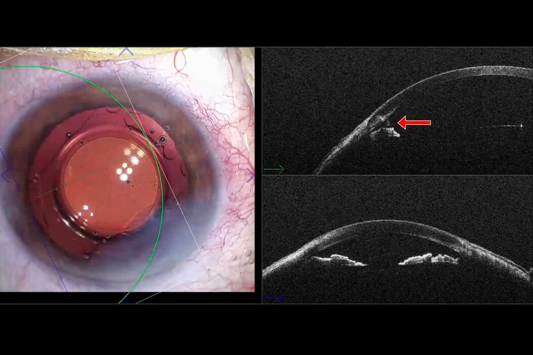
In this example, at the end of the surgery, I have a doubt about the presence of a residual fragment. Thanks to intraoperative OCT, I was able to highlight a residual fragment. It was quite a big fragment hidden behind the edema of the main incision.
[00:57 - 01:19]
In this second example, the smaller residual fragment was hidden behind the secondary incision. The problem is that corneal edema at the level of the incisions may reduce their visibility. That’s why using intraoperative OCT could be very useful.
[01:20 - 01:44]
What I usually do is to perform a 360° circular scan of the anterior segment. Using the microscope pedal, it is possible to go back over the sections to analyze the images. In this case, there was a residual fragment hidden behind the secondary incision edema.
[01:45 - 01:54]
And in this other case, there was a little residual fragment in the iridocorneal angle.
Benefits of intraoperative OCT
[01:55 - 02:26]
In my experience, intraoperative OCT could be very interesting in cataract surgery. We can visualize residual fragments; we can avoid readmission for secondary surgery a few days and weeks later, and we can reduce many postoperative complications. So, intraoperative OCT is a great tool to improve the quality and perfection of cataract surgery. Thank you for your attention.
Related Articles
-
RPE65 Gene Therapy Subretinal Injection: Benefits of Intraoperative OCT
Discover how RPE65 gene therapy subretinal injection procedures in patients with Leber congenital…
Feb 01, 2024Read article -

Dislocated Cataract Angle Closure Aided by Intraoperative OCT
Learn how a dislocated cataract was treated with angle closure assisted by intraoperative OCT to…
Dec 07, 2023Read article -

Glaucoma Stent Revision Surgery Guided by Intraoperative OCT
Learn about a glaucoma subconjunctival stent revision guided by intraoperative OCT and the important…
Nov 28, 2023Read article
