
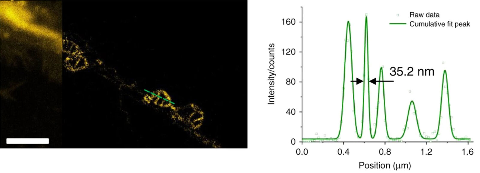
 and STED (right) imaging.")
Yang X., Yang Z., Wu Z., He Y., ShanC., Chai P., Ma C., Tian M., Teng J., Jin D., Yan W., Das P., Qu J. & Xi P.:
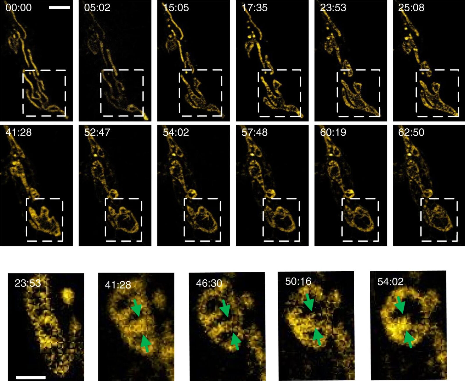
Mitochondrial dynamics quantitatively revealed by STED nanoscopy with an enhanced squaraine variant probe
Nature Communications volume 11, Article number: 3699 (2020)
Related Articles
-

Extended Live-cell Imaging at Nanoscale Resolution
Extended live-cell imaging with TauSTED Xtend. Combined spatial and lifetime information allow…
Mar 05, 2024Read article -
, actin network (ATTO 647N), and nuclear pore basket (CF 680R).")
The Guide to STED Sample Preparation
This guide is intended to help users optimize sample preparation for stimulated emission depletion…
Mar 05, 2024Read article -

Five-color FLIM-STED with One Depletion Laser
Webinar on five-color STED with a single depletion laser and fluorescence lifetime phasor…
Dec 14, 2022Read article
