
Multicolor Imaging with STELLARIS Confocal Platform
The STELLARIS family of confocal microscopes is equipped with next generation white light lasers (WLL). Paired with Power HyD detectors, STELLARIS provides complete spectral freedom. You can perform multicolor experiments to fit your research needs without being limited by the instrument. STELLARIS can also add an additional dimension to multicolor imaging by using the lifetime-based properties of fluorescent dyes/proteins with the TauSense set of tools.
Cytoskeleton and membranes in live cell imaged with TauSTED
Image acquired with STELLARIS STED.


TauSTED 775 resolves the intricate cytoskeleton network labeled with SiR-tubulin (glow - Spyrochrome), and trafficking vesicles labeled with CF594 (cyan - Biotium).
Endocytic pathway and mitochondria dynamics
Cytoskeleton and membranes in live cell imaged with TauSTED
Image acquired with STELLARIS STED. Live cell TauSTED on U2OS cells, using labels for actin (SiR-actin, glow), microtubules (SPY555-tubulin, cyan), and membranes (CF488A coupled to WGA, green). SiR and SPY are available from Spirochrome. CF dyes are available from Biotium Inc. Scale bar: 10 μm


Cytoskeleton and membranes in live cell imaged with TauSTED
Root-hypocotyl junction of Arabidopsis thaliana
Nature is multi-dimensional. To truly comprehend its complexity, we need to look in multiple dimensions. This detailed multicolor image of Arabadopsis thaliana root junction was obtained with just one click and one detector. With conventional confocal microscopy, it would be black and white. With STELLARIS what we see is transformed by an extra dimension of lifetime information. Not only is the remarkable architectural diversity of the actin-containing root system revealed with more clarity, but also its relationship to the cell wall and chloroplasts—structures that would be difficult or impossible to distinguish using the intensity data alone. The result is not just a snapshot of nature’s beauty, but an information-rich image that progresses our understanding of plant biology.

Cytoskeletal and mitochondrial interactions revealed
Most conventional confocal systems are equipped with only one or two discrete laser lines in the red. In addition, conventional GaAsP detectors are not sensitive in the NIR range. Both of these factors severely limit the number of red dyes that can be imaged in a single experiment with traditional confocal imaging systems. In the experiment shown here, Cos-7 cells were labeled with red-excitable probes specific for actin, mitochondrial outer membrane, and tubulin. Traditional confocal microscopy with a GaAsP detector detects only the SiRActin (Left). Using the extended WLL on STELLARIS 8 together with the HyD R detector, all 3 fluorescent probes were optimally excited and are visible in the image (Right). Imaging on STELLARIS not only made it possible to see the delicate interplay between mitochondria, actin and tubulin, but freed up the rest of the spectrum for additional fluorescent probes.
, AF750-Tom20 (760 – 790nm), AF790-Tubulin (810 – 850nm).")
Right: seen by STELLARIS 8 equipped with a Power HyD R detector. Spectral traces (right) show excitation (gray) and emission (red) spectra of fluorophores used in the experiment.
Sample courtesy: Dr.Jana Döhner and Dr.sc.nat. Urs Ziegler, University of Zürich.
Getting the most from live-cell experiments: fewer exposures, more information
When using traditional confocal microscopy to image several different fluorescent labels in the same sample, sequential imaging of each color channel is often needed to avoid spectral bleed-through, which can degrade image quality. In the case of a kinetic experiment, that means you may miss rapid dynamic events due to the increased time it takes to acquire each time point. In addition, your sample remains on the stage for longer, making it more challenging to maintain cell health for the duration the experiment. The image below was captured from live HeLa cells labeled with 4 different fluorophores to identify nuclei, actin, tubulin and plasma membrane. With STELLARIS, it was possible to collect all 4 channels in a single pass, rather than having to image the cells 4 times in succession.
, actin (yellow, SPY555), tublin (magenta, SPY650) and plasma membrane (gray, NIR750).")
to identify nuclei (cyan, Hoechst), actin (yellow, SPY555), tublin (magenta, SPY650) and plasma membrane (gray, NIR750).
LAS X Navigator – a GPS for your experiment
Intuitive sample navigation tools are essential to assure optimal setup of complex multicolor experiments and enable efficient exploration of large specimens. The LAS X Navigator is like a GPS for your experiment, helping you to quickly switch from searching image-by-image to seeing a full overview of your sample. With easy access to both the high-level overview and the minute details, the LAS X Navigator ensures that you always have a clear path to high-quality data.
Both the TLD and the TauContrast images were acquired with a 10x objective, and in each case a single tile is shown from the larger stitched image comprised of 201 tiles.

(shown on page X) labelled with Heidenhain azan trichrome stain. Transmitted
light detector (TLD) imaging was used to create an RGB overlay through excitation
with 488nm, 561nm and 638nm wavelengths.
Bottom: TauContrast image of the same field of view as above, maximum projection.
Discover at the speed of LIGHTNING
Confocal images obtained with STELLARIS contain more information than you see on the first glance. The LIGHTNING detection concept helps to uncover this hidden information from your specimen. With a single click and fully automatically, LIGHTNING extends the resolution of STELLARIS into super-resolution territory: resolve the fine structures and details, as small as 120 nm lateral resolution, which are elsewise simply not visible. Thanks to its deep integration and optimized GPU processing, LIGHTNING allows for the (near) real-time acquisition of super-resolution images with up to five color channels and a large field of view. Due to this flexibility, LIGHTNING works for any type of specimen and experiment.

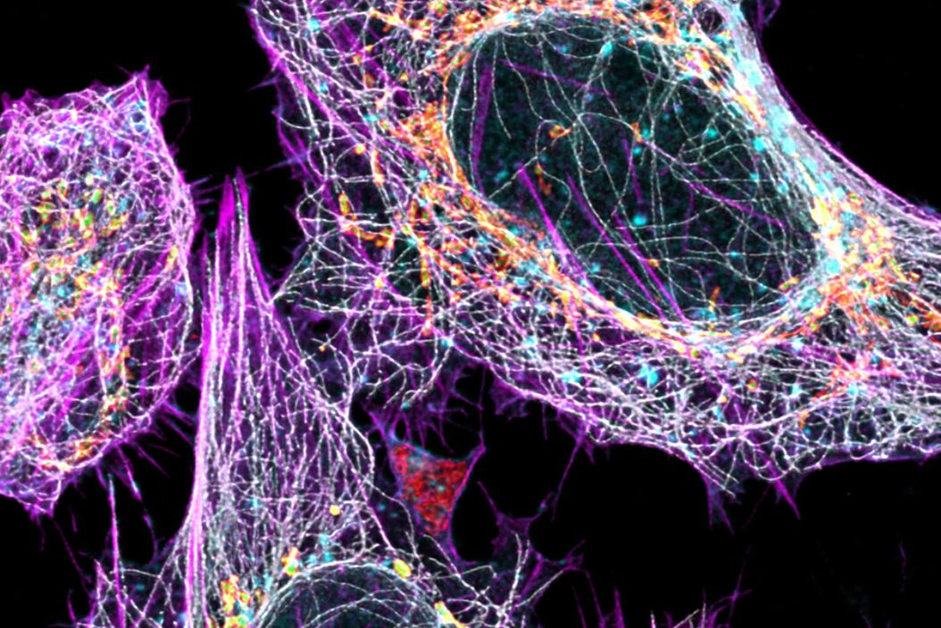
Overlay shows LIGHTNING data set labelled with AF488 (Tubulin, grey), SPY555 (Actin, magenta), MitoTrackerRed (Lumen of Mitochondria, green), Atto 647N (TOM 20, Mitochondria, red), CF770 WGA (Membranes, cyan).
Related Articles
-

Transforming Multiplexed 2D Data into Spatial Insights Guided by AI
Aivia 13 handles large 2D images and enables researchers to obtain deep insights into…
Dec 13, 2023Read article -

AI Microscopy Image Analysis – An Introduction
Artificial intelligence-guided microscopy image analysis and visualization is a powerful tool for…
Mar 24, 2022Read article -
Using Machine Learning in Microscopy Image Analysis
Recent exciting advances in microscopy technologies have led to exponential growth in quality and…
Jan 10, 2022Read article
