
Science Lab
Science Lab
The knowledge portal of Leica Microsystems offers scientific research and teaching material on the subjects of microscopy. The content is designed to support beginners, experienced practitioners and scientists alike in their everyday work and experiments. Explore interactive tutorials and application notes, discover the basics of microscopy as well as high-end technologies – become part of the Science Lab community and share your expertise!
Filter articles
Tags
Story Type
Products
Loading...
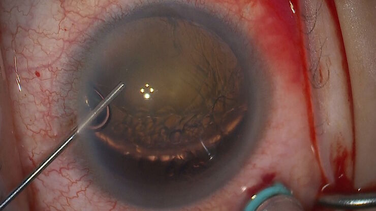
Dislocated Cataract Angle Closure Aided by Intraoperative OCT
Learn how a dislocated cataract was treated with angle closure assisted by intraoperative OCT to achieve long-term good results without future lens dislocation.
Loading...
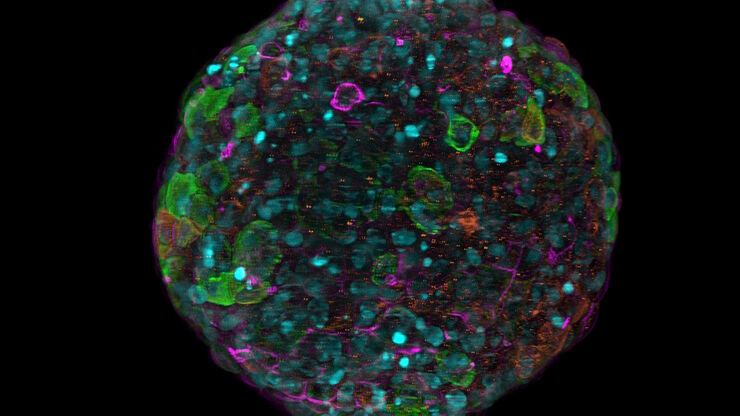
Notable AI-based Solutions for Phenotypic Drug Screening
Learn about notable optical microscope solutions for phenotypic drug screening using 3D-cell culture, both planning and execution, from this free, on-demand webinar.
Loading...
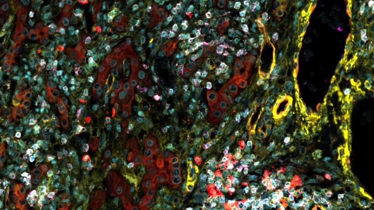
Understanding Tumor Heterogeneity with Protein Marker Imaging
Explore tumor heterogeneity and immune cell dynamics. See how quantitative imaging analysis reveals spatial relationships and molecular insights crucial for advancing cancer research and therapeutics.
Loading...
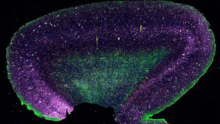
The Shape of the Brain: Spatial Biology of Alzheimer’s Disease
Uncover cell identity and brain structure in Alzheimer's disease with Cell DIVE multiplexed imaging, demonstrating how spatial biology can lead to advances in therapy development for…
Loading...
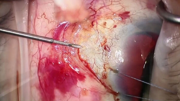
Glaucoma Stent Revision Surgery Guided by Intraoperative OCT
Learn about a glaucoma subconjunctival stent revision guided by intraoperative OCT and the important role it plays to ensure the best outcome.
Loading...
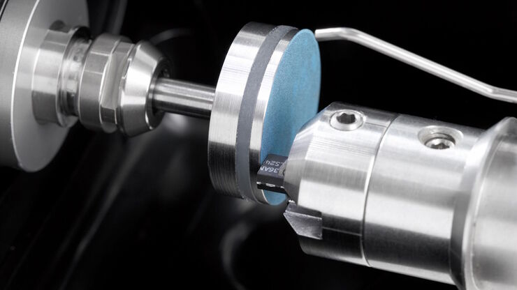
Cross-section Analysis for Electronics Manufacturing
This article describes cross-section analysis for electronics concerning quality control and failure analysis of printed circuit boards (PCBs) and assemblies (PCBAs), integrated circuits (ICs), etc.
Loading...
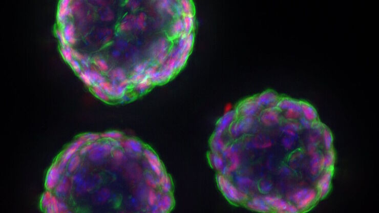
How to Get Deeper Insights into your Organoid and Spheroid Models
In this eBook, learn about key considerations for imaging 3D cultures, such as organoids and spheroids, and discover microscopy solutions to shed new insights into dynamic processes in 3D real-time
Loading...
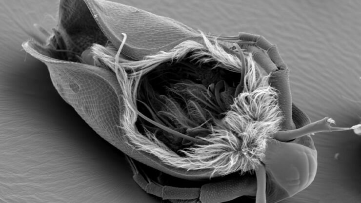
Streamline your EM Sample Preparation Workflow for Biological Applications
Master EM sample preparation, including ultramicrotomy, for life sciences in this expert eBook!
Loading...
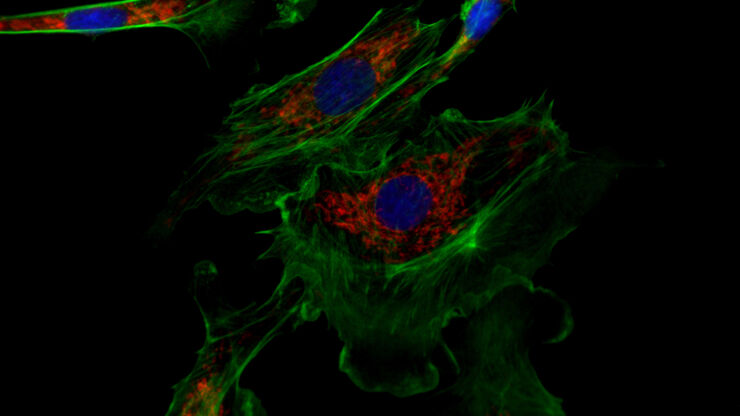
Studying Virus Replication with Fluorescence Microscopy
The results from research on SARS-CoV-2 virus replication kinetics, adaption capabilities, and cytopathology in Vero E6 cells, done with the help of fluorescence microscopy, are described in this…