
Science Lab
Science Lab
The knowledge portal of Leica Microsystems offers scientific research and teaching material on the subjects of microscopy. The content is designed to support beginners, experienced practitioners and scientists alike in their everyday work and experiments. Explore interactive tutorials and application notes, discover the basics of microscopy as well as high-end technologies – become part of the Science Lab community and share your expertise!
Filter articles
Tags
Story Type
Products
Loading...
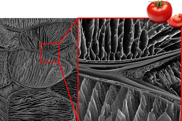
Studying the Microstructure of Natural Polymers in Fine Detail
The potential of cryogenic broad ion beam milling used in combination with scanning electron microscopy (cryo-BIB-SEM) for imaging and analyzing the microstructure of cryogenically stabilized soft…
Loading...

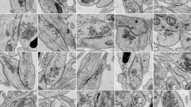
Introduction to Ultramicrotomy
When studying samples, to visualize their fine structure with nanometer scale resolution, most often electron microscopy is used. There are 2 types: scanning electron microscopy (SEM) which images the…
Loading...
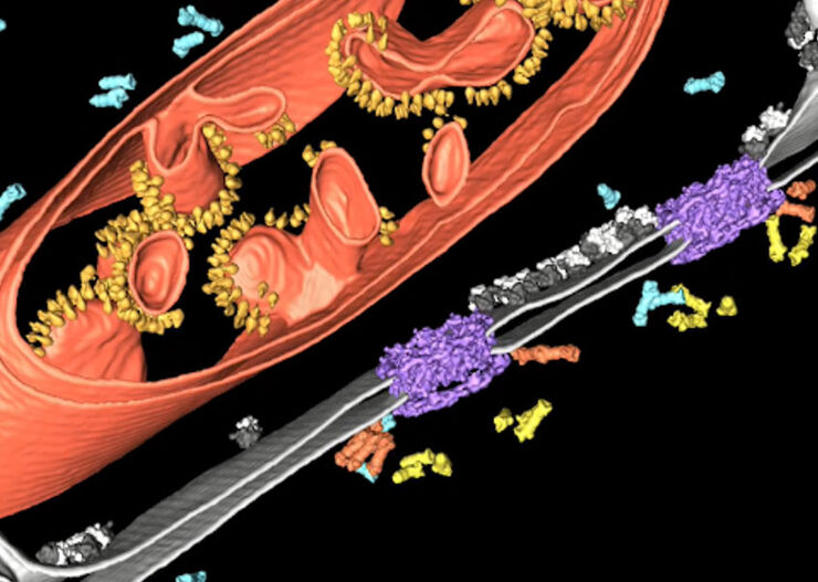
Improve Cryo Electron Tomography Workflow
Leica Microsystems and Thermo Fisher Scientific have collaborated to create a fully integrated cryo-tomography workflow that responds to these research needs: Reveal cellular mechanisms at…
Loading...

Development of Fluorescence Lifetime Imaging Microscopy (FLIM) and its Relevance for Functional Imaging
Prof. Ammasi Periasamy, Director, Keck Center for Cellular Imaging, University of Virginia, was interviewed by Dr. Giulia Ossato, Product Manager functional imaging, during Leica Microsystems Meets…
Loading...

How to Adapt Grain Size Analysis of Metallic Alloys to Your Needs
Metallic alloys, such as steel and aluminum, have an important role in a variety of industries, including automotive and transportation. In this report, the importance of grain size analysis for alloy…
Loading...
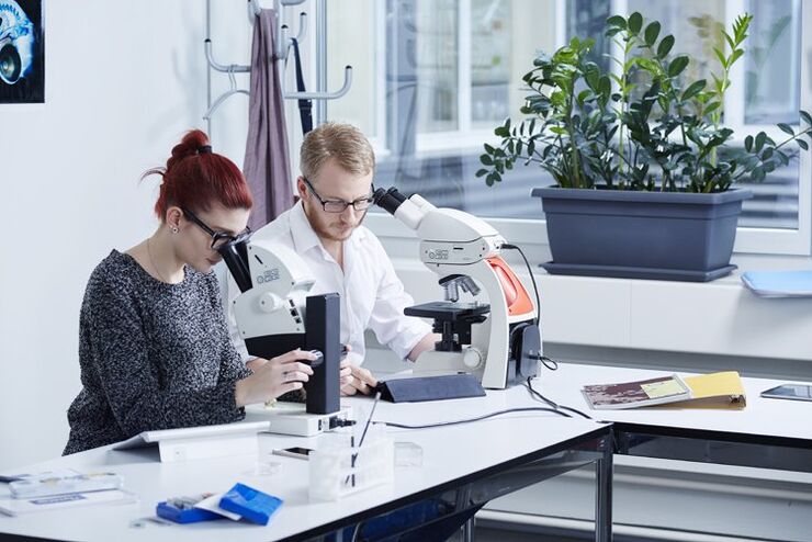
Digital Classroom Options
As teachers, you know your big challenge is to catch and keep the students’ attention and the best chance for this is by making the environment interactive. In the case of the Microscopy Classroom, we…
Loading...
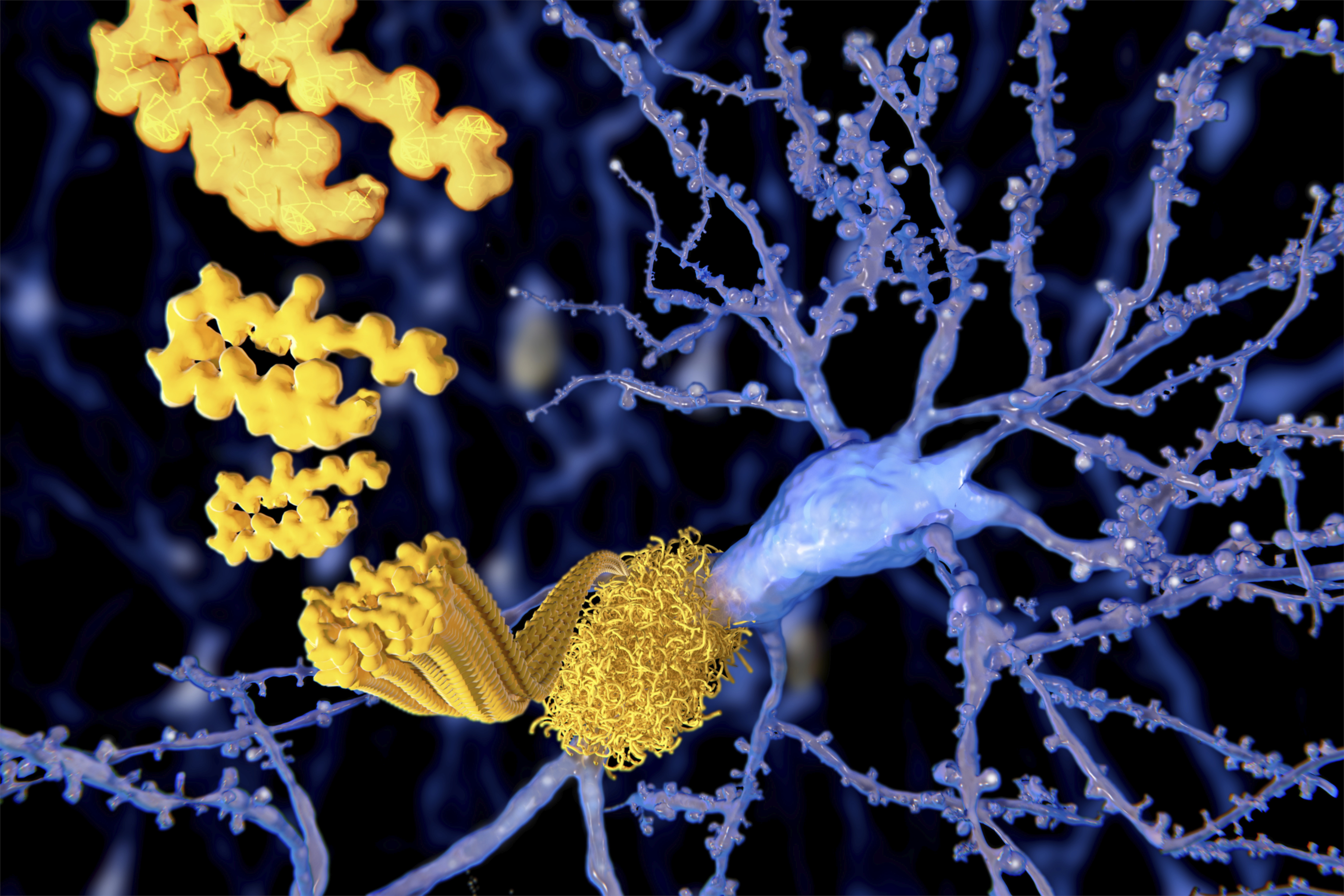
Alzheimer Plaques: fast Visualization in Thick Sections
More than 60% of all diagnosed cases of dementia are attributed to Alzheimer’s disease. Typical of this disease are histological alterations in the brain tissue. So far, there is no cure for this…
Loading...

Expert Knowledge on High Pressure Freezing and Freeze Fracturing in the Cryo SEM Workflow
Get an insight in the working methods of the laboratory and learn about the advantages of Cryo SEM investigation in EM Sample Preparation. Find out how high pressure freezing, freeze fracturing and…
Loading...
Bridging Structure and Dynamics at the Nanoscale through Optogenetics and Electrical Stimulation
Nanoscale ultrastructural information is typically obtained by means of static imaging of a fixed and processed specimen. However, this is only a snapshot of one moment within a dynamic system in…