
Science Lab
Science Lab
The knowledge portal of Leica Microsystems offers scientific research and teaching material on the subjects of microscopy. The content is designed to support beginners, experienced practitioners and scientists alike in their everyday work and experiments. Explore interactive tutorials and application notes, discover the basics of microscopy as well as high-end technologies – become part of the Science Lab community and share your expertise!
Filter articles
Tags
Story Type
Products
Loading...
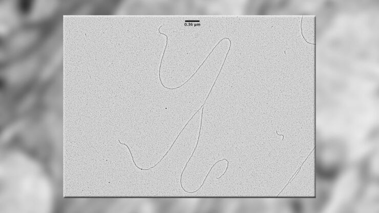
Visualization of DNA Molecules
Precise low angle rotary shadowing with heavy metals (like platinum) can be used in transmission electron microscopy (TEM) to observe molecular details of objects previously absorbed on a thin, low…
Loading...
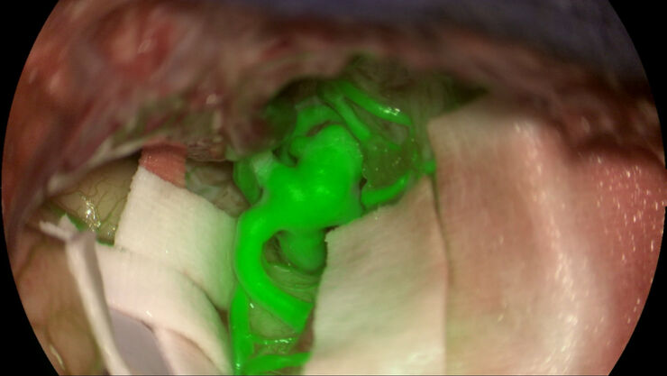
The Future of Fluorescence in Vascular Neurosurgery
In vascular neurosurgery, surgical microscopes are used to provide a magnified and illuminated view of the surgical field. Although surgeons benefit greatly from the superb image quality and optical…
Loading...

Using U-Shaped Glass Capillaries for Sample Mounting
The DLS microscope system from Leica Microsystems is an innovative concept which integrates the Light Sheet Microscopy technology into the confocal platform. Due to its unique optical architecture,…
Loading...
The Fundamentals and History of Fluorescence and Quantum Dots
At some point in your research and science career, you will no doubt come across fluorescence microscopy. This ubiquitous technique has transformed the way in which microscopists can image, tag and…
Loading...

Multiphoton Microscopy – a Satisfied Wish List
With the variable beam expander (VBE) from Leica Microsystems, we can look deeper into microscopic objects. The user can tune the beam to attain the deepest insight and finest detail
Loading...
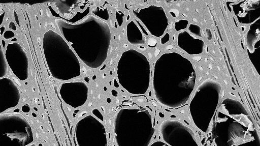
Practical Applications of Broad Ion Beam Milling
Mechanical polishing can be time consuming and frustrating. It can also introduce unwanted artifacts when preparing cross-sectioned samples for electron backscatter diffraction (EBSD) in the scanning…
Loading...

Digital Microscopy in Earth Science
Classical polarized light (compound) microscopes can only be used for prepared samples, because the working distance they offer is insufficient for whole samples. This means that thicker and bigger…
Loading...

Eyepieces, Objectives and Optical Aberrations
This article covers the components of the eyepieces and how to adjust them correctly to suit your eyes.
Loading...

Mission Impossible Accomplished: Tunable Colors for Non-descanning Detection
Leica Microsystems’ 4Tune detector, the key component of the SP8 DIVE Deep In Vivo Explorer, provides spectrally tunable image recording with non-descanning detection. An innovative solution for…