
Science Lab
Science Lab
The knowledge portal of Leica Microsystems offers scientific research and teaching material on the subjects of microscopy. The content is designed to support beginners, experienced practitioners and scientists alike in their everyday work and experiments. Explore interactive tutorials and application notes, discover the basics of microscopy as well as high-end technologies – become part of the Science Lab community and share your expertise!
Filter articles
Tags
Story Type
Products
Loading...

Introduction to Widefield Microscopy
This article gives an introduction to widefield microscopy, one of the most basic and commonly used microscopy techniques. It also shows the basic differences between widefield and confocal…
Loading...
Optimization of the Interplay of Optical Components for Aberration Free Microscopy
Optical microscopes are used to magnify objects which are otherwise invisible for the human eye. For this purpose high quality optics is necessary to achieve appropriate resolution. However, besides…
Loading...

About the Most Important Considerations When Imaging Deep Into Mouse Tissue
When operating a confocal microscope, or when discussing features and parameters of such a device, we inescapably mention the pinhole and its diameter. This short introductory document is meant to…
Loading...
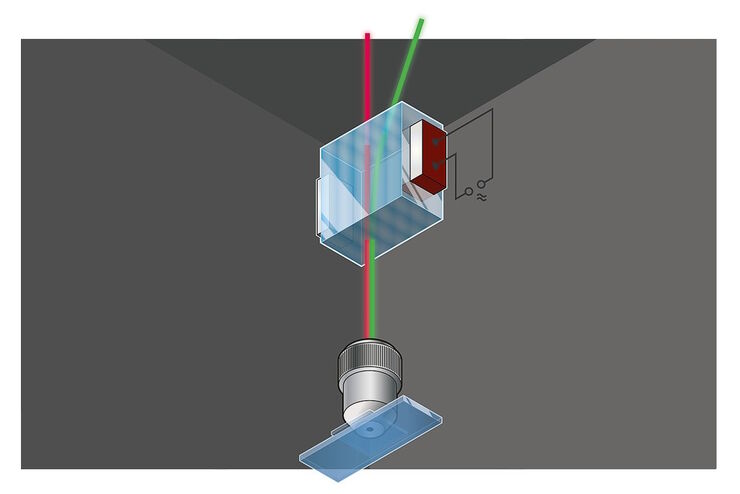
Primary Beam Splitting Devices for Confocal Microscopes
Current fluorescence microscopy employs incident illumination which requires separation of illumination and emission light. The classical device performing this separation is a color-dependent beam…
Loading...

Photoactivatable, Photoconvertible, and Photoswitchable Fluorescent Proteins
Fluorescent proteins (FPs) such as GFP, YFP or DsRed are powerful tools to visualize cellular components in living cells. Nevertheless, there are circumstances when classical FPs reach their limits.…
Loading...

Pinhole Effect in Confocal Microscopes
When operating a confocal microscope, or when discussing features and parameters of such a device, we inescapably mention the pinhole and its diameter. This short introductory document is meant to…
Loading...
 stereo microscope for a task like surgery.")
Rodent and Small-Animal Surgery
Learn how you can perform rodent (mouse, rat, hamster) and small-animal surgery efficiently with a microscope for developmental biology and medical research applications by reading this article.
Loading...

Navigating Through the Brain
One of the challenges of neurosurgery is orientation at the surgical site. When resecting tumors, removing arteriovenous malformations, or clipping aneurysms, surgeons often have to work near healthy…
Loading...
 illuminated with wide-band UV excitation. Note the tissue structure with blue autofluorescence. Right: Same tissue and same immunostaining with FITC label illuminated with epi-illumination using narrow")
Milestones in Incident Light Fluorescence Microscopy
Since the middle of the last century, fluorescence microscopy developed into a bio scientific tool with one of the biggest impacts on our understanding of life. Watching cells and proteins with the…