
Science Lab
Science Lab
The knowledge portal of Leica Microsystems offers scientific research and teaching material on the subjects of microscopy. The content is designed to support beginners, experienced practitioners and scientists alike in their everyday work and experiments. Explore interactive tutorials and application notes, discover the basics of microscopy as well as high-end technologies – become part of the Science Lab community and share your expertise!
Filter articles
Tags
Story Type
Products
Loading...

Imaging and Analyzing Zebrafish, Medaka, and Xenopus
Discover how to image and analyze zebrafish, medaka, and Xenopus frog model organisms efficiently with a microscope for developmental biology applications from this article.
Loading...
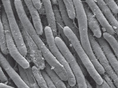
Bacteria Protocol - Critical Point Drying of E. coli for SEM
Application Note for Leica EM CPD300 - Critical point drying of E. coli with subsequent platinum / palladium coating and SEM analysis. Sample was inserted into a filter disc (Pore size: 16 - 40 μm)…
Loading...
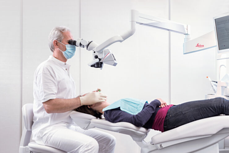
Successful Endodontic Treatment with Dental Operating Microscopes
In endodontics, accurate treatment is not only dependent on the technical skills and knowledge of the dentist, but also on clear, detailed visualization of the surgical field. As the outcome of an…
Loading...

Investigating Fruit Flies (Drosophila melanogaster)
Learn how to image and investigate Drosophila fruit fly model organisms efficiently with a microscope for developmental biology applications from this article.
Loading...

Studying Caenorhabditis elegans (C. elegans)
Find out how you can image and study C. elegans roundworm model organisms efficiently with a microscope for developmental biology applications from this article.
Loading...
BABB Clearing and Imaging for High Resolution Confocal Microscopy
Multipohoton microscopy experiment using Leica TCS SP8 MP and Leica 20x/0.95 NA BABB immersion objective.
Understanding kidney microanatomy is key to detecting and identifying early events in kidney…
Loading...
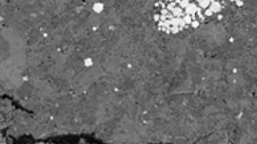
Ion Beam Polishing of Sample Surfaces - Sample Preparation for SEM
Application Note for Leica EM RES102 - Ion milling can be used to reduce the roughness of sample surfaces. Small angles less than 6° with respect to the sample surface are necessary. The high voltage…
Loading...
")
Epoxy Resin Embedding of Animal and Human Tissues for Pathological Diagnosis and Research
Application Note for Leica EM AMW - The tissues were fixed in the modified Karnovsky fixative generally by immersion overnight (at minimum 4h at room temperature). Then pieces of approx. 1mm3 were cut…
Loading...
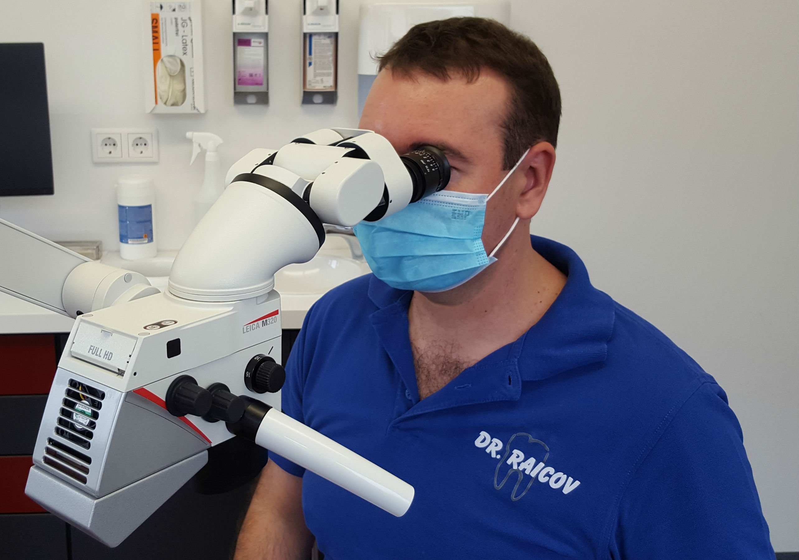
The Dental Microscope in Endodontics
In dentistry a bright, magnified view into deep cavities supports detailed diagnosis and precise therapy, particularly in the field of endodontics. In this interview Dr. Dean Raicov explains the…