
Science Lab
Science Lab
The knowledge portal of Leica Microsystems offers scientific research and teaching material on the subjects of microscopy. The content is designed to support beginners, experienced practitioners and scientists alike in their everyday work and experiments. Explore interactive tutorials and application notes, discover the basics of microscopy as well as high-end technologies – become part of the Science Lab community and share your expertise!
Filter articles
Tags
Story Type
Products
Loading...
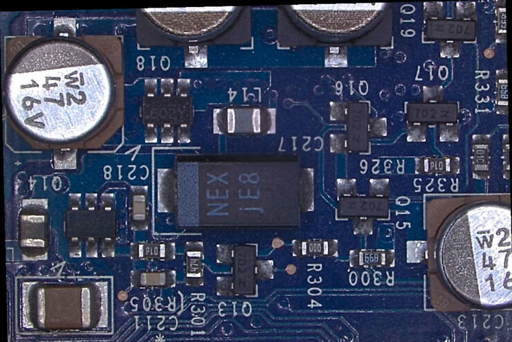
Rapid and Reliable Examination of PCBs & PCBAs with Digital Microscopy
Digital microscopes provide users with a convenient and rapid way to acquire high-quality, reliable image data and make quick inspection and analysis of printed circuit boards (PCBs) and assemblies…
Loading...
. Courtesy: Thomas Mathivet, PhD")
Windows on Neurovascular Pathologies
Discover how innate immunity can sustain deleterious effects following neurovascular pathologies and the technological developments enabling longitudinal studies into these events.
Loading...

Ophthalmology Case Study: Corneal Transplantation
Learn about the use of intraoperative Optical Coherence Tomography in Corneal Transplantation and how it helps achieve correct positioning of donor tissue.
Loading...

What is the FusionOptics Technology?
Leica stereo microscopes with FusionOptics provide optimal 3D perception. The brain merges two images, one with large depth of field and the other with high resolution, into one 3D image.
Loading...

Microscope Ergonomics
This article explains microscope ergonomics and how it helps users work in comfort, enabling consistency and efficiency. Learn how to set up the workplace to keep good posture when using a microscope.
Loading...
 and labelled with MitoTracker Green.")
The Power of Reproducibility, Collaboration and New Imaging Technologies
In this webinar you willl learn what impacts reproducibility in microscopy, what resources and initiatives there are to improve education and rigor and reproducibility in microscopy and how…
Loading...

Examining Developmental Processes In Cancer Organoids
Interview: Prof. Bausch and Dr. Pastucha, Technical University of Munich, discuss using microscopy to study development of organoids, stem cells, and other relevant disease models for biomedical…
Loading...
. Image courtesy of Prof. Hui Guo, School of Life Sciences, Central South University, China")
How Microscopy Helps the Study of Mechanoceptive and Synaptic Pathways
In this podcast, Dr Langenhan explains how microscopy helps his team to study mechanoceptive and synaptic pathways, their challenges, and how they overcome them.
Loading...

3D, AR & VR for Teaching in Neurosurgery
Discover the evolution of neurosurgical teaching and how 3D, Augmented Reality and Virtual Reality can help better learn anatomy and acquire surgical skills.