
Science Lab
Science Lab
The knowledge portal of Leica Microsystems offers scientific research and teaching material on the subjects of microscopy. The content is designed to support beginners, experienced practitioners and scientists alike in their everyday work and experiments. Explore interactive tutorials and application notes, discover the basics of microscopy as well as high-end technologies – become part of the Science Lab community and share your expertise!
Filter articles
Tags
Story Type
Products
Loading...

What are the Challenges in Neuroscience Microscopy?
eBook outlining the visualization of the nervous system using different types of microscopy techniques and methods to address questions in neuroscience.
Loading...

Central Nervous System (CNS) Development and Activity in Organisms
This article shows how studying central nervous system (CNS) development in Drosophila-melanogaster embryos expressing a GCaMP calcium indicator in the neurons can be improved with a THUNDER Imager.
Loading...
Going Beyond Deconvolution
Widefield fluorescence microscopy is often used to visualize structures in life science specimens and obtain useful information. With the use of fluorescent proteins or dyes, discrete specimen…
Loading...

Diseases Linked to Scaffold Proteins and Signaling
This article shows how diseases related to scaffold proteins and protein signaling can be studied in zebrafish models efficiently with a THUNDER Imager.
Loading...
 and THUNDER image (bottom).")
The Neural Crest (NC)
This article discusses how the study of neural crest (NC) development in chicken embryos is aided with haze-free imaging using a THUNDER Imager 3D Assay. Proper specification, migration, and…
Loading...

Fast, High Acuity Imaging and AI-assisted Analysis
The use of state-of-the-art AI systems is pushing image analysis into a new generation. Challenges like the conflict between imaging power and sample integrity are being overcome with THUNDER’s…
Loading...

Create New Options for Live Cell Imaging
The use of state-of-the-art AI systems is pushing image analysis into a new generation. Challenges like the conflict between imaging power and sample integrity are being overcome with THUNDER’s…
Loading...
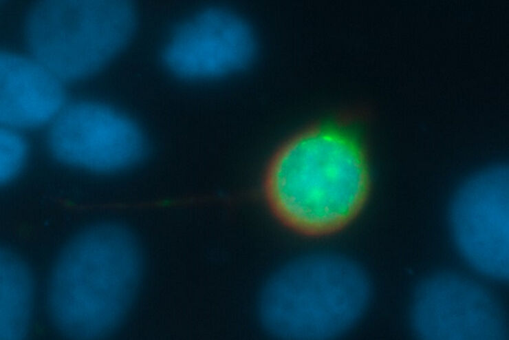
 expressed in the sensory neurons.")
Fast, High-contrast 3D Imaging of Sensory Neurons
This article discusses how fast, high-contrast 3D imaging of dorsal root ganglion (DRG) tissue with a THUNDER Imager Tissue using large volume computational clearing (LVCC) allows sensory neurons to…
Loading...

Visualizing Retinal Interactions to Study Eye Diseases
This article shows how interactions between endothelial cells, blood vessels, microglia, and astrocytes in mouse retina can be studied efficiently with a THUNDER Imager 3D Cell Culture and Large…