
Science Lab
Science Lab
The knowledge portal of Leica Microsystems offers scientific research and teaching material on the subjects of microscopy. The content is designed to support beginners, experienced practitioners and scientists alike in their everyday work and experiments. Explore interactive tutorials and application notes, discover the basics of microscopy as well as high-end technologies – become part of the Science Lab community and share your expertise!
Filter articles
Tags
Story Type
Products
Loading...

Download EM Workflow Solutions Booklet
This publication is a compilation of appropriate workflows for the most frequently used sample preparation methods, like Correlative Methodologies, Optogenetics & Electro-Physiology, Surface Analysis,…
Loading...
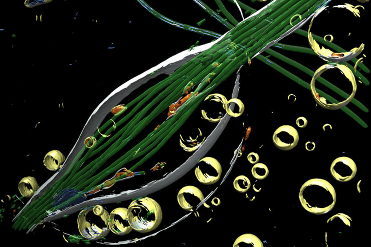
Exploring the Structure and Life Cycle of Viruses
The SARS-CoV-2 outbreak started in late December 2019 and has since reached a global pandemic, leading to a worldwide battle against COVID-19. The ever-evolving electron microscopy methods offer a…
Loading...
Fast, High-quality Vitrification with the EM ICE High Pressure Freezer
The EM ICE High Pressure Freezer was developed with a unique freezing principle and uses only a single pressurization and cooling liquid: liquified nitrogen (LN2). This design enables three major…
Loading...

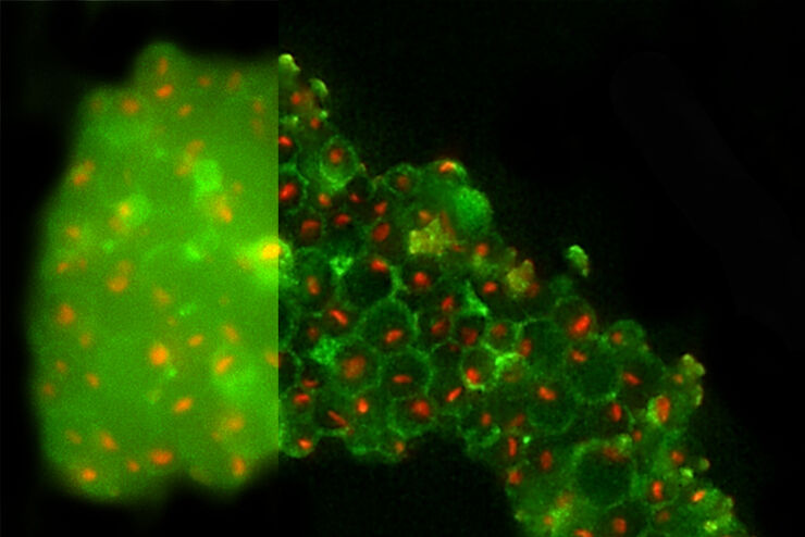
Targeting Active Recycling Nuclear Pore Complexes using Cryo Confocal Microscopy
In this article, how cryo light microscopy and, in particular cryo confocal microscopy, is used to improve the reliability of cryo EM workflows is described. The quality of the EM grids and samples is…
Loading...
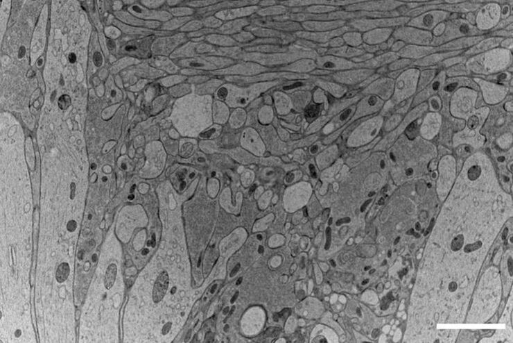
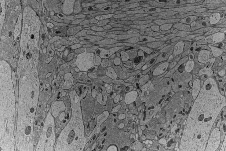
Investigating Synapses in Brain Slices with Enhanced Functional Electron Microscopy
A fundamental question of neuroscience is: what is the relationship between structural and functional properties of synapses? Over the last few decades, electrophysiology has shed light on synaptic…
Loading...
Advancing Cell Biology with Cryo-Correlative Microscopy
Correlative light and electron microscopy (CLEM) advances biological discoveries by merging different microscopes and imaging modalities to study systems in 4D. Combining fluorescence microscopy with…
Loading...
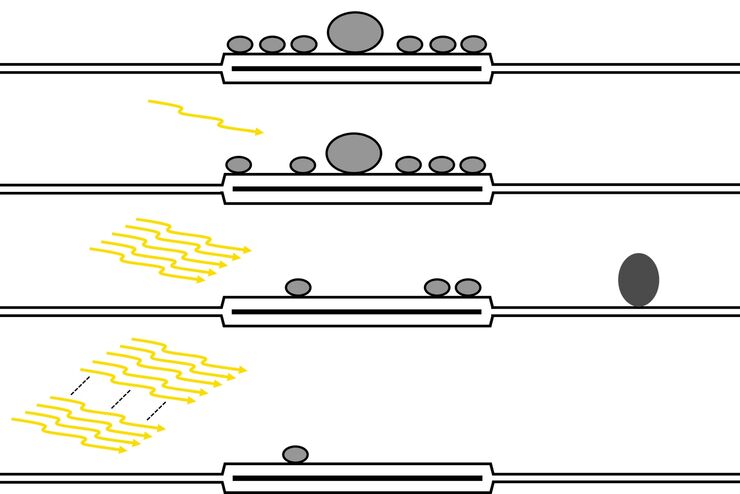
High-pressure freezing: Revealing functional mechanisms of synaptic transmission
Learn more about applying optogenetic stimulation in the EM ICE and how this technology has the potential to reveal structural and functional mechanisms of synaptic transmission. Get a detailed…
Loading...
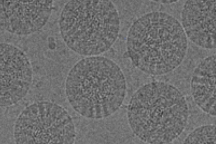
Workflows and Instrumentation for Cryo-electron Microscopy
Cryo-electron microscopy is an increasingly popular modality to study the structures of macromolecular complexes and has enabled numerous new insights in cell biology. In recent years, cryo-electron…
Loading...
Crystal Clear Cryo Light-microscopy Images
This article describes how computational clearing of cryo light microscopy images improves the identification of cellular targets for cryo electron-microscopy.