
Science Lab
Science Lab
The knowledge portal of Leica Microsystems offers scientific research and teaching material on the subjects of microscopy. The content is designed to support beginners, experienced practitioners and scientists alike in their everyday work and experiments. Explore interactive tutorials and application notes, discover the basics of microscopy as well as high-end technologies – become part of the Science Lab community and share your expertise!
Filter articles
Tags
Story Type
Products
Loading...

Technical Terms for Digital Microscope Cameras and Image Analysis
Learn more about the basic principles behind digital microscope camera technologies, how digital cameras work, and take advantage of a reference list of technical terms from this article.
Loading...

Epi-Illumination Fluorescence and Reflection-Contrast Microscopy
This article discusses the development of epi-illumination and reflection contrast for fluorescence microscopy concerning life-science applications. Much was done by the Ploem research group…
Loading...

Life Science Research: Which Microscope Camera is Right for You?
Deciding which microscope camera best fits your experimental needs can be daunting. This guide presents the key factors to consider when selecting the right camera for your life science research.
Loading...

Examining Developmental Processes In Cancer Organoids
Interview: Prof. Bausch and Dr. Pastucha, Technical University of Munich, discuss using microscopy to study development of organoids, stem cells, and other relevant disease models for biomedical…
Loading...

Central Nervous System (CNS) Development and Activity in Organisms
This article shows how studying central nervous system (CNS) development in Drosophila-melanogaster embryos expressing a GCaMP calcium indicator in the neurons can be improved with a THUNDER Imager.
Loading...

How to Image Histological and Fluorescent Samples with One System
VIDEO ON DEMAND - How to image histological and fluorescent samples with one system. FluoSync, the new technology embedded into Mica enables the imaging of both histological staining and fluorescence…
Loading...
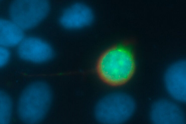
Going Beyond Deconvolution
Widefield fluorescence microscopy is often used to visualize structures in life science specimens and obtain useful information. With the use of fluorescent proteins or dyes, discrete specimen…
Loading...
How to Radically Simplify Workflows in Your Imaging Facility
VIDEO ON DEMAND - How to radically simplify imaging workflows and generate meaningful results with less time and effort using a highly automated microscope that unites widefield and confocal imaging.
Loading...

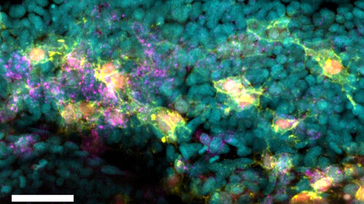
Diseases Linked to Scaffold Proteins and Signaling
This article shows how diseases related to scaffold proteins and protein signaling can be studied in zebrafish models efficiently with a THUNDER Imager.