
Science Lab
Science Lab
The knowledge portal of Leica Microsystems offers scientific research and teaching material on the subjects of microscopy. The content is designed to support beginners, experienced practitioners and scientists alike in their everyday work and experiments. Explore interactive tutorials and application notes, discover the basics of microscopy as well as high-end technologies – become part of the Science Lab community and share your expertise!
Filter articles
Tags
Story Type
Products
Loading...
 and mito OM (red) in a live U2OS cell")
Multicolor 4D Super Resolution Light Sheet Microscopy
The AI Microscopy Symposium offers a unique forum for discussing the latest AI-based technologies and tools in the field of microscopy and biomedical imaging. In this scientific presentation, Yuxuan…
Loading...
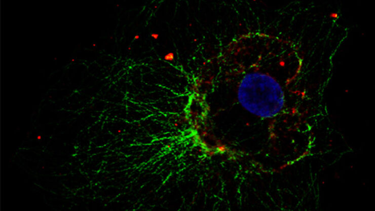
How to Prepare your Specimen for Immunofluorescence Microscopy
Immunofluorescence (IF) is a powerful method for visualizing intracellular processes, conditions and structures. IF preparations can be analyzed by various microscopy techniques (e.g. CLSM,…
Loading...
Live-Cell Imaging Techniques
The understanding of complex and/or fast cellular dynamics is an important step for exploring biological processes. Therefore, today’s life science research is increasingly focused on dynamic…
Loading...

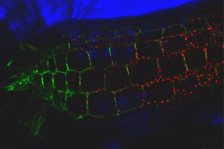
Imaging of Anti-Cancer Drug Uptake in Spheroids using DLS
Spheroid 3D cell culture models mimic the physiology and functions of living tissues making them a useful tool to study tumor morphology and screen anti-cancer drugs. The drug AZD2014 is a recognized…
Loading...

Intravital Microscopy of Cancer
Join our guest speaker Prof Dr Jacco van Rheenen, as he presents his work on the identity, behavior and fate of cells that drive the initiation and progression of cancer.
Loading...
Save Time and Effort with AI-assisted Fluorescence Image Analysis
The powerful synergy of THUNDER and Aivia analyze fluorescence images with greater accuracy, even when using low light excitation.
Loading...
Tracking Single Cells Using Deep Learning
AI-based solutions continue to gain ground in the field of microscopy. From automated object classification to virtual staining, machine and deep learning technologies are powering scientific…
Loading...

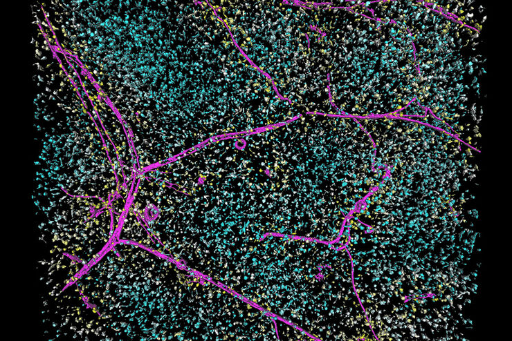
Learning the Cellular Architecture from its Optical Properties
In the last 3 years, microscopists have started to use "AI based" solutions for a wide range of applications, including image acquisition optimization (smart microscopy), object classification, image…
Loading...
 in 3D")
Artificial Intelligence and Confocal Microscopy – What You Need to Know
This list of frequently asked questions provides “hands-on” answers and is a supplement to the introductory article about Dynamic Signal Enhancement powered by Aivia "How Artificial Intelligence…