
Science Lab
Science Lab
The knowledge portal of Leica Microsystems offers scientific research and teaching material on the subjects of microscopy. The content is designed to support beginners, experienced practitioners and scientists alike in their everyday work and experiments. Explore interactive tutorials and application notes, discover the basics of microscopy as well as high-end technologies – become part of the Science Lab community and share your expertise!
Filter articles
Tags
Story Type
Products
Loading...
, the cis-golgi matrix protein GM130 (AF488, green), and the trans-golgi network membrane protein TGN46 (AF647, red).")
Golgi Organizational Changes in Response to Cell Stress
VIDEO ON DEMAND - In this episode of MicaCam, our special guest George Galea from EMBL Heidelberg will look at HeLa Kyoto cells treated with various chemotherapeutic agents to investigate their effect…
Loading...

Multiplexed Imaging Types, Benefits and Applications
Multiplexed imaging is an emerging and exciting way to extract information from human tissue samples by visualizing many more biomarkers than traditional microscopy. By observing many biomarkers…
Loading...

3D Tissue Imaging: From Fast Overview To High Resolution With One Click
3D Tissue imaging is a widespread discipline in the life sciences. Researchers use it to reveal detailed information of tissue composition and integrity, to make conclusions from experimental…
Loading...

How To Perform Fast & Stable Multicolor Live-Cell Imaging
With the help of live-cell imaging researchers gain insights into dynamic processes of living cells up to whole organisms. This includes intracellular as well as intercellular activities. Protein or…
Loading...

Imaging of Cardiac Tissue Regeneration in Zebrafish
Learn how to image cardiac tissue regeneration in zebrafish focusing on cell proliferation and response during recovery. MicaCam Episode 04 with Laura Peces-Barba Castaño from the Max Planck…
Loading...

Multiplexed Imaging Glossary
Human tissues contain a many different biomarkers which can be explored to reveal the workings of biological pathways and interrogate disease states. Recent technological advances have made it…
Loading...
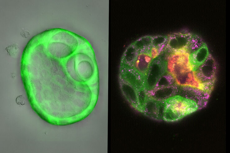
How Does The Cytoskeleton Transport Molecules?
VIDEO ON DEMAND - See how 3D cysts derived from MDCK cells help scientists understand how proteins are transported and recycled in tissues and the role of the cytoskeleton in this transport.
Loading...
 embryo, from sphere stage to somite stages.")
Studying Early Phase Development of Zebrafish Embryos
VIDEO ON DEMAND - This second edition of MicaCam focuses on combining widefield and confocal imaging to study the early-stage development of zebrafish embryos (Danio rerio), from oocyte to…
Loading...

Tumor MHC Expression and Intralesional IL2 Response in Melanoma
Genomics profiling and Cell DIVE multiplex imaging allows researchers to understand the immune cell phenotypes that most strongly predict response to IL2 immunotherapy in melanoma patients suffering…