
Science Lab
Science Lab
The knowledge portal of Leica Microsystems offers scientific research and teaching material on the subjects of microscopy. The content is designed to support beginners, experienced practitioners and scientists alike in their everyday work and experiments. Explore interactive tutorials and application notes, discover the basics of microscopy as well as high-end technologies – become part of the Science Lab community and share your expertise!
Filter articles
Tags
Story Type
Products
Loading...

Every Clue Counts – Forensics Inconceivable Without Microscopy
There is no crime without clues. They may be obvious, like a cartridge case at the scene of the crime or clear signs of crowbar damage on a door. But sometimes, clues are microscopically small.…
Loading...
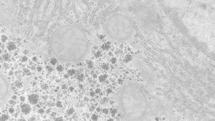
Perusing Alternatives for Automated Staining of TEM Thin Sections
Contrast in transmission electron microscopy (TEM) is mainly produced by electron scattering at the specimen: Structures that strongly scatter electrons are referred to as electron dense and appear as…
Loading...

50 Years of Image Analysis
Modern image analysis systems perform highly sophisticated image processing functions on images from an automated microscope and digital camera. 50 years ago, the first image analysis system was…
Loading...
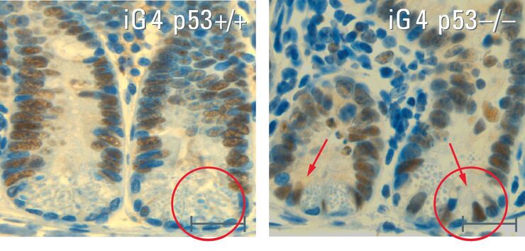
Stem Cells in the Aging Process – the Signal Protein p53 as "Guardian of the Genome"
The physiological process of aging is an elementary part of life. The processes responsible for the aging process and the effect they have is one of the key focuses of biomedical research. Dr. Yvonne…
Loading...

Nobel Prize 2012 in Physiology or Medicine for Stem Cell Research
The Nobel Prize recognizes two scientists who discovered that mature, specialised cells can be reprogrammed to become immature cells capable of developing into all tissues of the body. Their findings…
Loading...

CARS Microscopy: Imaging Characteristic Vibrational Contrast of Molecules
Coherent anti-Stokes Raman scattering (CARS) microscopy is a technique that generates images based on the vibrational signatures of molecules. This imaging methods does not require labeling, yet…
Loading...

Fluorescent Proteins - From the Beginnings to the Nobel Prize
Fluorescent proteins are the fundament of recent fluorescence microscopy and its modern applications. Their discovery and consequent development was one of the most exciting innovations for life…
Loading...
Super-Resolution GSDIM Microscopy
The nanoscopic technique GSDIM (ground state depletion microscopy followed by individual molecule return) provides a detailed image of the spatial arrangement of proteins and other biomolecules within…
Loading...
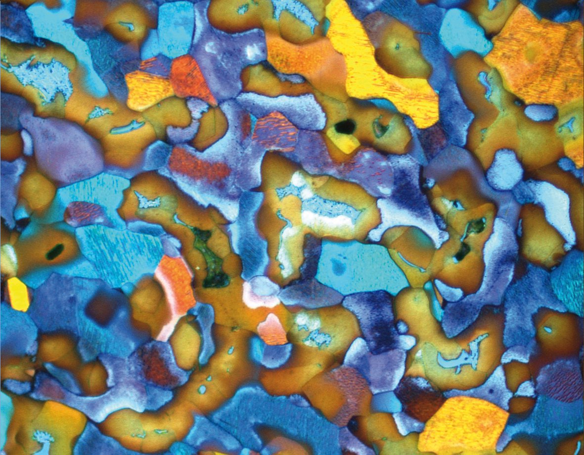
Metallography with Color and Contrast
The examination of microstructure morphology plays a decisive role in materials science and failure analysis. There are many possibilities of visualizing the real structures of materials in the light…