
Science Lab
Science Lab
The knowledge portal of Leica Microsystems offers scientific research and teaching material on the subjects of microscopy. The content is designed to support beginners, experienced practitioners and scientists alike in their everyday work and experiments. Explore interactive tutorials and application notes, discover the basics of microscopy as well as high-end technologies – become part of the Science Lab community and share your expertise!
Filter articles
Tags
Story Type
Products
Loading...

Studying Ocular Birth Defects
This article discusses how lens formation and ocular birth defects can be studied with sharp widefield microscopy images which are acquired rapidly. The mouse ocular lens is used as a model to study…
Loading...

Studying Wound Healing of Smooth Muscle Cells
This article discusses how wound healing of cultured smooth muscle cells (SMCs) in multiwell plates can be reliably studied over time with less effort using a specially configured Leica inverted…
Loading...

How AR Helps in the Surgical Treatment of Moyamoya Disease
Moyamoya disease is a rare chronic occlusive cerebrovascular disorder characterized by progressive stenosis in the terminal portion of the internal carotid artery and an abnormal vascular network at…
Loading...

Multiplex Imaging Reveals Survival Markers after Cancer Care
Colorectal cancer is a high incidence and high mortality cancer. Currently, postoperative chemotherapy benefits only a minority of patients, and thus, new tools are necessary to screen patients and…
Loading...

Using GLOW800 AR in Radial Forearm Flap Free Phalloplasty
In this video, Chief Microsurgeon Professor Küntscher and his team perform a radial forearm free flap phalloplasty and use ICG fluorescence imaging to show the blood flow in the whole flap from the…
Loading...
 expressed in the sensory neurons.")
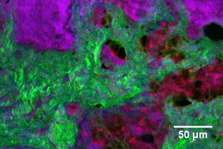
Fast, High-contrast 3D Imaging of Sensory Neurons
This article discusses how fast, high-contrast 3D imaging of dorsal root ganglion (DRG) tissue with a THUNDER Imager Tissue using large volume computational clearing (LVCC) allows sensory neurons to…
Loading...
 or Minor’s syndrome")
Minor’s Syndrome Surgical Intervention by Prof. Vincent Darrouzet
Minor’s disease, also called Superior Semicircular Canal Dehiscence (SSCD) or Minor’s syndrome, is a rare disorder of the inner ear that affects hearing and balance. The disease is characterized by…
Loading...
 and THUNDER (right) image of Ewing Sarcoma cells (SK-ES-1)")
Visualizing the Mitotic Spindle in Cancer Cells
This article demonstrates how this research is aided by visualizing more details of mitotic spindles in Ewing Sarcoma cells using the THUNDER Imager Tissue and Large Volume Computational Clearing…
Loading...
Formulated Product Characterization with SRS Microscopy
Creams, pastes, gels, emulsions, and tablets are ubiquitous across a wide range of manufacturing sectors from pharmaceuticals and consumer health products to agrochemicals and paint. To improve…