
Science Lab
Science Lab
The knowledge portal of Leica Microsystems offers scientific research and teaching material on the subjects of microscopy. The content is designed to support beginners, experienced practitioners and scientists alike in their everyday work and experiments. Explore interactive tutorials and application notes, discover the basics of microscopy as well as high-end technologies – become part of the Science Lab community and share your expertise!
Filter articles
Tags
Story Type
Products
Loading...

What are the Challenges in Neuroscience Microscopy?
eBook outlining the visualization of the nervous system using different types of microscopy techniques and methods to address questions in neuroscience.
Loading...

How is Microscopy Used in Spatial Biology? A Microscopy Guide
Different spatial biology methods in microscopy, such as multiplex imaging, are helping to better understand tissue landscapes. Learn more in this microscopy guide.
Loading...

Why 3D Microscope Visualization Enriches Surgical ENT Education
Learn about the benefits of using a Leica surgical microscope with 3D visualization for surgical ENT education.
Loading...
FluoSync - a Fast & Gentle Method for Unmixing Multicolour Images
In this white paper, we focus on a fast and reliable method for obtaining high-quality multiplex images in fluorescence microscopy. FluoSync combines an existing method for hybrid unmixing with…
Loading...
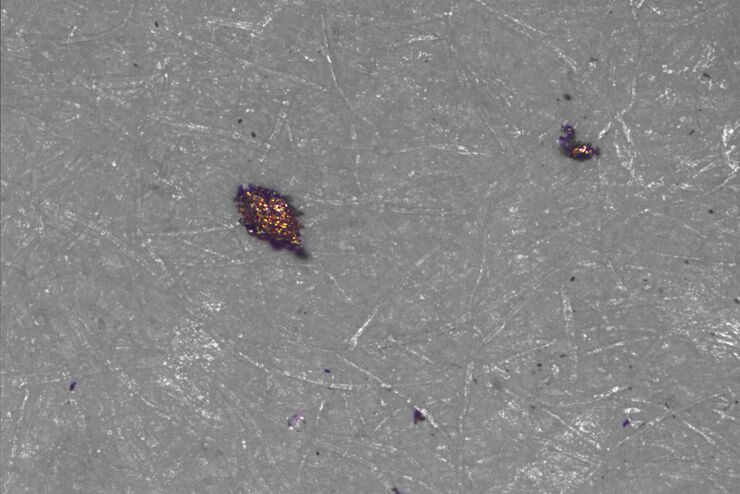
Factors to Consider for a Cleanliness Analysis Solution
Choosing the right cleanliness analysis solution is important for optimal quality control. This article discusses the important factors that should be taken into account to find the solution that best…
Loading...

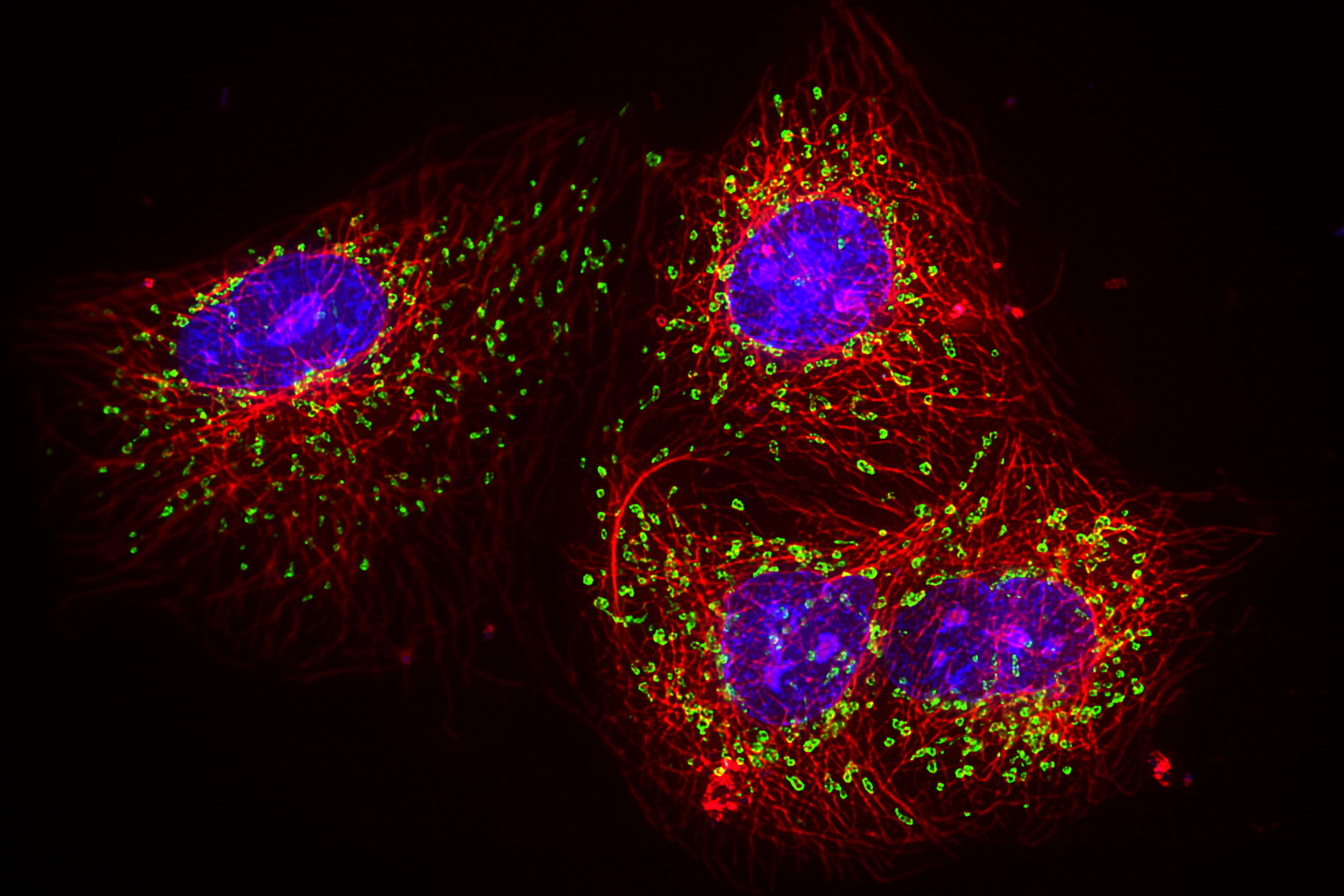
New Imaging Tools for Cryo-Light Microscopy
New cryo-light microscopy techniques like LIGHTNING and TauSense fluorescence lifetime-based tools reveal structures for cryo-electron microscopy.
Loading...

Fast, High Acuity Imaging and AI-assisted Analysis
The use of state-of-the-art AI systems is pushing image analysis into a new generation. Challenges like the conflict between imaging power and sample integrity are being overcome with THUNDER’s…
Loading...

Create New Options for Live Cell Imaging
The use of state-of-the-art AI systems is pushing image analysis into a new generation. Challenges like the conflict between imaging power and sample integrity are being overcome with THUNDER’s…
Loading...

How to Target Fluorescent Structures in 3D for Cryo-FIB Milling
This article describes the major steps of the cryo-electron tomography workflow including super-resolution cryo-confocal microscopy. We describe how subcellular structures can be precisely located in…