
Science Lab
Science Lab
The knowledge portal of Leica Microsystems offers scientific research and teaching material on the subjects of microscopy. The content is designed to support beginners, experienced practitioners and scientists alike in their everyday work and experiments. Explore interactive tutorials and application notes, discover the basics of microscopy as well as high-end technologies – become part of the Science Lab community and share your expertise!
Filter articles
Tags
Story Type
Products
Loading...

Create New Options for Live Cell Imaging
The use of state-of-the-art AI systems is pushing image analysis into a new generation. Challenges like the conflict between imaging power and sample integrity are being overcome with THUNDER’s…
Loading...
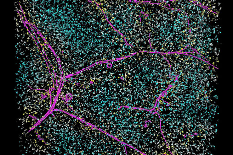
 expressed in the sensory neurons.")
Fast, High-contrast 3D Imaging of Sensory Neurons
This article discusses how fast, high-contrast 3D imaging of dorsal root ganglion (DRG) tissue with a THUNDER Imager Tissue using large volume computational clearing (LVCC) allows sensory neurons to…
Loading...

Find Relevant Specimen Details from Overviews
Switch from searching image by image to seeing the full overview of samples quickly and identifying the important specimen details instantly with confocal microscopy. Use that knowledge to set up…
Loading...

Accurately Analyze Fluorescent Widefield Images
The specificity of fluorescence microscopy allows researchers to accurately observe and analyze biological processes and structures quickly and easily, even when using thick or large samples. However,…
Loading...
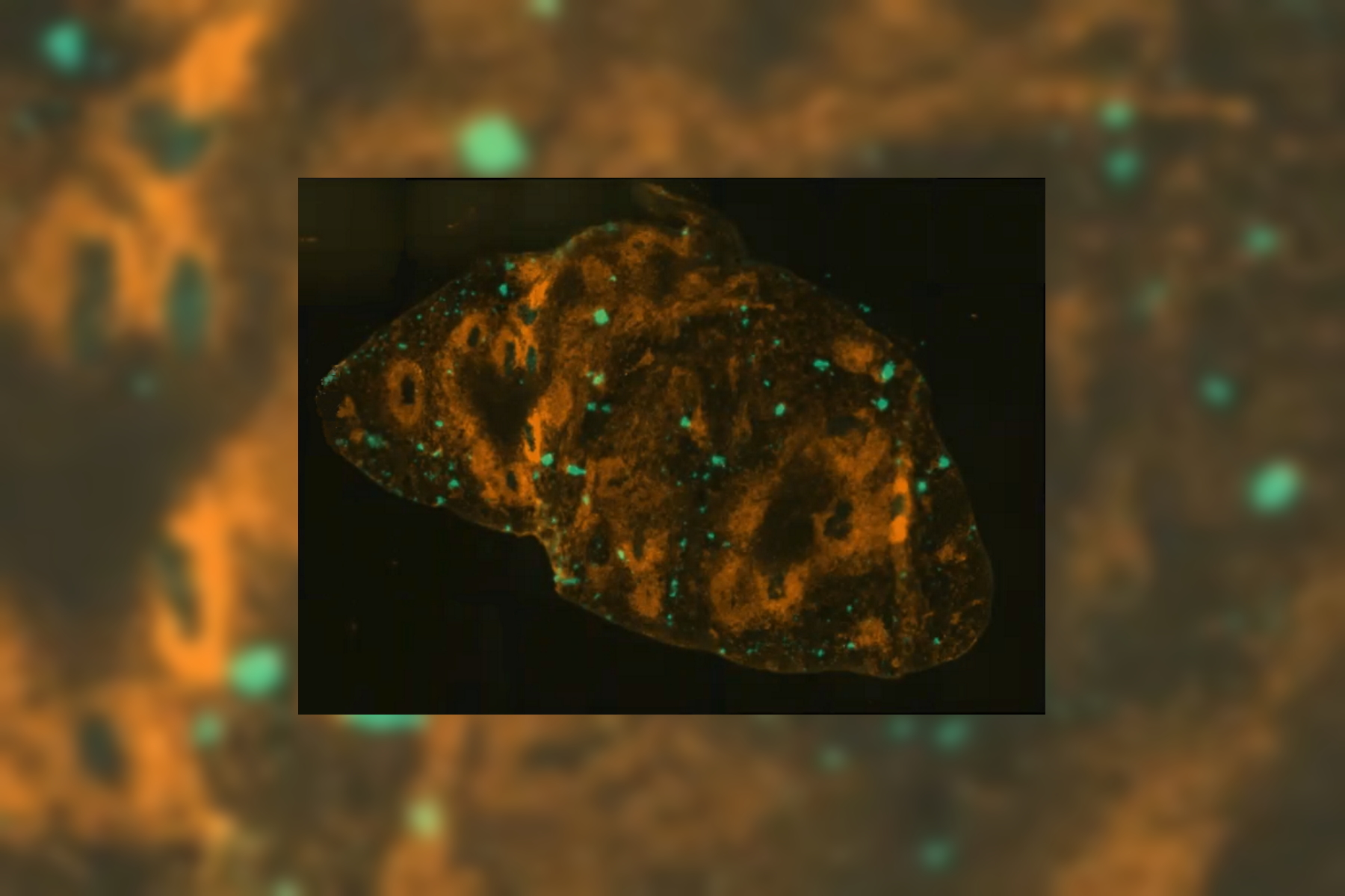
 and THUNDER (right) image of Ewing Sarcoma cells (SK-ES-1)")
Visualizing the Mitotic Spindle in Cancer Cells
This article demonstrates how this research is aided by visualizing more details of mitotic spindles in Ewing Sarcoma cells using the THUNDER Imager Tissue and Large Volume Computational Clearing…
Loading...
Save Time and Effort with AI-assisted Fluorescence Image Analysis
The powerful synergy of THUNDER and Aivia analyze fluorescence images with greater accuracy, even when using low light excitation.
Loading...

High-resolution 3D Imaging to Investigate Tissue Ageing
Award-winning researcher Dr. Anjali Kusumbe demonstrates age-related changes in vascular microenvironments through single-cell resolution 3D imaging of young and aged organs.
Loading...

How to Improve Live Cell Imaging with Coral Life
For live-cell CLEM applications, light microscopy imaging is a critical step for identifying the right cell in the right state at the right time. In this article, Leica experts share their insights on…
Loading...
Optimizing THUNDER Platform for High-Content Slide Scanning
With rising demand for full-tissue imaging and the need for FL signal quantitation in diverse biological specimens, the limits on HC imaging technology are tested, while user trainability and…