Cryo CLEM from Leica Microsystems
The motto „From Eye to Insight” of Leica Microsystems states it correctly: as humans we are mainly driven by our visual system. What we can see, we can easily believe. A picture is worth a thousand words. To get pictures from life, or better scientific samples, microscopes are the choice as soon as the sample, or its interesting content is leaving the cm range. Thus, it is simply too tiny to be visualized by the naked eye.
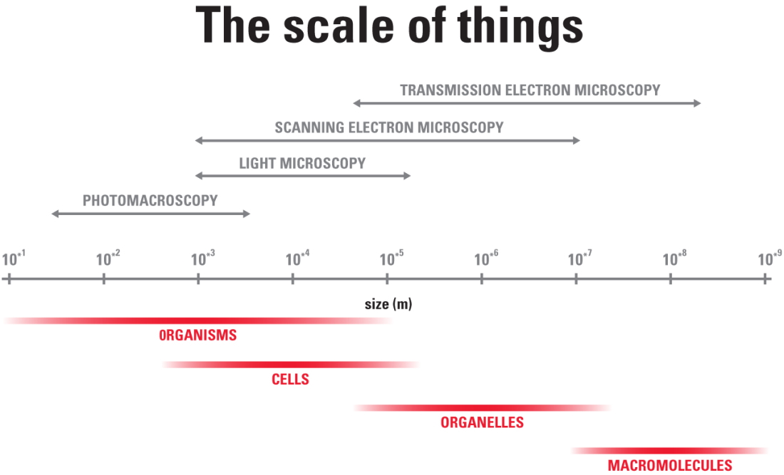
Microscope technology
There are different types of microscopes which are helpful for specific samples in specific dimension ranges. Light microscopes range from stereo microscopes, macroscopes, fluorescence microscopes, confocal microscopes with many tricks to increase resolution, contrast, etc. However, all of these light microscopes have their limits when it comes to magnification. On this border electron microscopy steps in which allows to see even smaller structures which are restricted for light microscopes. The limits of both worlds, light and electron microscopy have been pushed in the past to new limits and will be pushed further in the future.
Sample preparation
Aside from the microscope technology, the sample preparation plays a crucial role for the visualization. Scientists want to see the native state of their sample or at least to get as close as possible. Chemical fixation techniques can alter the native state. Living cells are so far restricted for light microscopic investigations. Preservation of the native state for electron microscopy can be achieved by complete vitrification of the sample.
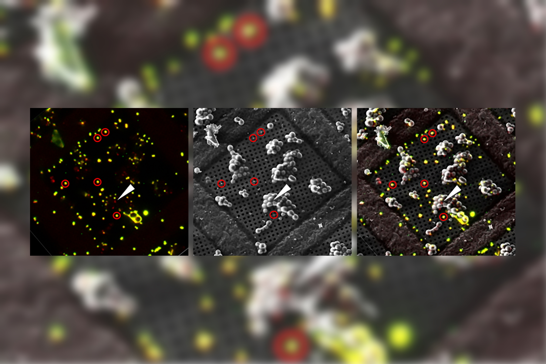
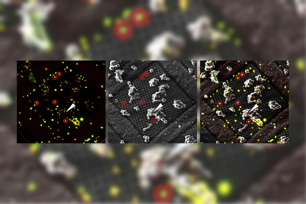
CLEM solution
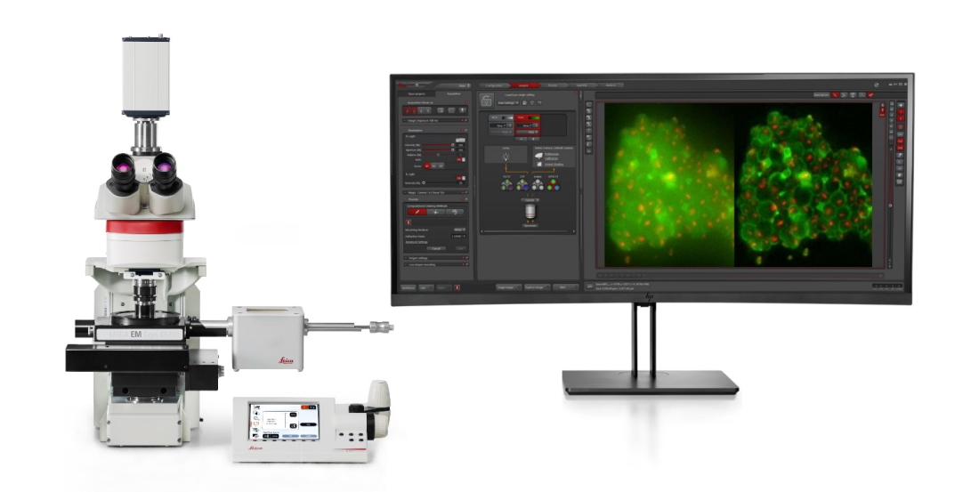
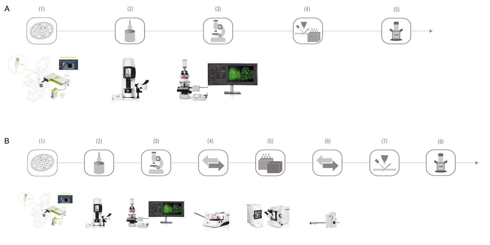
At Leica Microsystems we continuously improve the combination of both light and electron microscopy. This process is commonly known as CLEM (correlative light & electron microscopy). Beside the established tools for EM samples preparation like the grid plunger Leica EM GP2, the high-pressure freezer Leica EM ICE, the vacuum transfer system Leica EM VCT and the new Nano workflow combining the Leica THUNDER imager live cell and the Leica EM ICE to preserve a sample by vitrification at a specific time point, the Leica THUNDER imager EM CryoCLEM is the key tool for the CLEM approaches under cryogenic conditions.
The joint forces of EM sample preparation knowledge and scientific microscope expertise allow Leica Microsystems to provide this solution from a single supplier. The Leica CryoCLEM solution enabled since its launch many high impact publications:
Strnad et al., Sci Rep. 2015 Dec 10;5:18029. doi: 10.1038/srep18029. [1]
Schorb et al., J Struct Biol. 2017 Feb;197(2):83-93. doi: 10.1016/j.jsb.2016.06.020. [2]
Kolovou et al., Methods Cell Biol. 2017;140:85-103. doi: 10.1016/bs.mcb.2017.03.011. [3]
Schorb & Sieckmann, Methods Cell Biol. 2017;140:321-333. doi: 10.1016/bs.mcb.2017.03.012. [4]
Yang et al., J Struct Biol. 2021 Feb 18;213(2):107709. doi: 10.1016/j.jsb.2021.107709. [5]
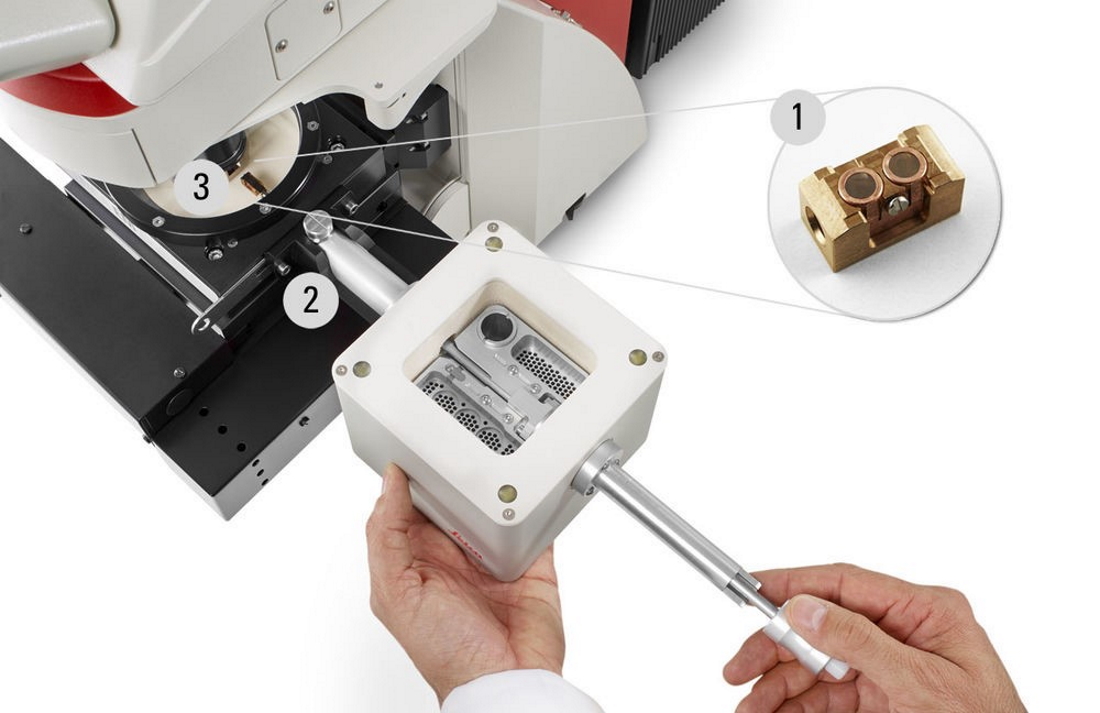
The key of the Leica Cryo CLEM solution is the transfer shuttle which enables a safe transfer of the sample under cryogenic conditions.
THUNDER Imager EM Cryo CLEM - Media | Products | Leica Microsystems (leica-microsystems.com)
The unique shuttle transfer design allows us to separate the sample transfer system and the microscope stage. This minimizes cross contaminations. Time savings are an additional effect as the shuttle can be smoothly dried during fluorescent imaging or simply a second shuttle can be prepared for sample transfer while imaging takes place.
The separation of the fluorescent imaging and the FIB milling bares many advantages. For example, fluorescent imaging allows a quick quality check before the sample goes into the cryo FIB-SEM or Cryo TEM. This maximizes the usage of FIB-SEM and TEM system time as the suitability of the sample is given and proofed upfront. Before the sample makes its way to the EM world, fluorescent expression of targets can be confirmed, grid damages can be inspected, etc. As most EM systems are available in facilities, light microscopy proven high-quality samples allow to maximize the result during booked sessions.
Solution evolution
Based on the initial combination of a widefield fluorescence microscope with the special cryo stage to allow the sample to stay under cryogenic/vitrified conditions after plunge freezing, many solution approaches have been performed. The combination with Leica’s computational clearing method THUNDER or even with confocal microscope including LIGHTNING are commercially available. In addition, scientists work on the combination with super resolution technologies: Wolff et al., Biol Cell. 2016 Sep;108(9):245-58. doi: 10.1111/boc.201600008 [6]. Cryo-FIB Lift out is on the roadmap for further developments as well: Klumpe et al., bioRxiv preprint doi: https://doi.org/10.1101/2021.05.19.444745 [7].
Practical application
Along with the excitement of technology development and pushing the methodologic limits, cryo CLEM approaches led to scientific insights. Structural biology is clearly the most important field for cryo CLEM:
Paul et al., J Cell Biol. 2020 Sep 7;219(9):e201911154 [8] uncovered an unexpected versatility in cytoskeletal form, more specifically they discovered filamentous actin (F-actin) inside the microtubule lumen through in situ cryo-electron tomography (cryo-ET) analysis of these small molecule–induced projection. Therefore, cryo fluorescence microscopy helped to identify cells with emerging projections.
Allegretti et al., Nature. 2020 Oct;586(7831):796-800 [9] were able to directly identify structure-function relationships of nuclear pore complexes (NPCs) in their cellular context on the macromolecular level. Cryo CLEM investigations helped here to identify nitrogen starvation levels and therefore timepoints of autophagy machinery recruitment to the NPC.
Li et al., Prog Biophys Mol Biol. 2021 Mar;160:87-96 [10] used cryo fluorescent microscopy to identify and mark mitochondria in outgrowing neurites of PC12 cells. The fluorescent markings served for easy navigation for the following cryo electrotomography (cryo ET).
Klein et al., Commun Biol. 2021 Jan 29;4(1):137 [11] investigated lamellar bodies (LBs) and found an undescribed outer membrane dome protein complex (OMDP) to be limiting membrane of LBs. Their data achieved by the combination of fluorescent microscopy of labelled LBs with FIB-milling and Cryo ET plus back correlation to Cryo fluorescent microscopy suggest that LB biogenesis is driven by parallel membrane sheet import and by the curvature of the limiting membrane to maximize lipid storage capacity.
In general, cryo CLEM is a valid workflow approach to study host-pathogen interaction, especially virus infections simply due to the size of viruses. Examples for such approaches are:
Strnad et al., Sci Rep. 2015 Dec 10;5:18029. doi: 10.1038/srep18029. [1]
Outlook
As cryo CLEM is a rather young technology which evolved in the past year to a tool which is not restricted to experts any longer, it will be a key step of future scientific workflows to uncover the unseen. Dr. Julia König, Product Manager of the Leica THUNDER Imager EM Cryo CLEM summarizes:
So far, we've uncovered the secrets inside cells; what will we discover inside whole tissues?
References
- Strnad M, Elsterová J, Schrenková J, Vancová M, Rego RO, Grubhoffer L, Nebesářová, Correlative cryo-fluorescence and cryo-scanning electron microscopy as a straightforward tool to study host-pathogen interactions, J.Sci Rep. 2015 Dec 10;5:18029. doi: 10.1038/srep18029.
- Schorb M, Gaechter L, Avinoam O, Sieckmann F, Clarke M, Bebeacua C, Bykov YS, Sonnen AF, Lihl R, Briggs JAG, New hardware and workflows for semi-automated correlative cryo-fluorescence and cryo-electron microscopy/tomography, J Struct Biol. 2017 Feb;197(2):83-93. doi: 10.1016/j.jsb.2016.06.020. Epub 2016 Jun 28.
- Kolovou A, Schorb M, Tarafder A, Sachse C, Schwab Y, Santarella-Mellwig R, A new method for cryo-sectioning cell monolayers using a correlative workflow, Methods Cell Biol. 2017;140:85-103. doi: 10.1016/bs.mcb.2017.03.011. Epub 2017 Apr 13.
- Schorb M, Sieckmann F, Matrix MAPS-an intuitive software to acquire, analyze, and annotate light microscopy data for CLEM, Methods Cell Biol. 2017;140:321-333. doi: 10.1016/bs.mcb.2017.03.012. Epub 2017 Apr 19.
- Yang JE, Larson MR, Sibert BS, Shrum S, Wright ER, CorRelator: Interactive software for real-time high precision cryo-correlative light and electron microscopy, J Struct Biol. 2021 Feb 18;213(2):107709. doi: 10.1016/j.jsb.2021.107709. Online ahead of print.
- Wolff G, Hagen C, Grünewald K, Kaufmann R, Towards correlative super-resolution fluorescence and electron cryo-microscopy, Biol Cell. 2016 Sep;108(9):245-58. doi: 10.1111/boc.201600008. Epub 2016 Jun 22.
- Klumpe S, Fung HKH, Goetz SK, Zagoriy I, Hampoelz B, Zhang X, Erdmann PS, Baumbach J, Müller CW, Beck M, Plitzko JM, Mahamid J, A Modular Platform for Streamlining Automated Cryo-FIB Workflows, bioRxiv preprint doi: https://doi.org/10.1101/2021.05.19.444745
- Paul DM, Mantell J, Borucu U, Coombs J, Surridge KJ, Squire JM, Verkade P, Dodding MP, In situ cryo-electron tomography reveals filamentous actin within the microtubule lumen, J Cell Biol. 2020 Sep 7;219(9):e201911154. doi: 10.1083/jcb.201911154.
- Allegretti M, Zimmerli CE, Rantos V, Wilfling F, Ronchi P, Fung HKH, Lee CW, Hagen W, Turoňová B, Karius K, Börmel M, Zhang X, Müller CW, Schwab Y, Mahamid J, Pfander B, Kosinski J, Beck M, In-cell architecture of the nuclear pore and snapshots of its turnover, Nature. 2020 Oct;586(7831):796-800. doi: 10.1038/s41586-020-2670-5. Epub 2020 Sep 2.
- Li X, Park D, Chang Y, Radhakrishnan A, Wu H, Wang P, Liu J, A mammalian system for high-resolution imaging of intact cells by cryo-electron tomography, Prog Biophys Mol Biol. 2021 Mar;160:87-96. doi: 10.1016/j.pbiomolbio.2020.09.005. Epub 2020 Oct 13.
- Klein S, Wimmer BH, Winter SL, Kolovou A, Laketa V, Chlanda P, Post-correlation on-lamella cryo-CLEM reveals the membrane architecture of lamellar bodies, Commun Biol. 2021 Jan 29;4(1):137. doi: 10.1038/s42003-020-01567-z.
- Hampton CM, Strauss JD, Ke Z, Dillard RS, Hammonds JE, Alonas E, Desai TM, Marin M, Storms RE, Leon F, Melikyan GB, Santangelo PJ, Spearman PW, Wright ER, Correlated fluorescence microscopy and cryo-electron tomography of virus-infected or transfected mammalian cells, Nat Protoc. 2017 Jan;12(1):150-167. doi: 10.1038/nprot.2016.168. Epub 2016 Dec 15.
- Vancová M, Rudenko N, Vaněček J, Golovchenko M, Strnad M, Rego ROM, Tichá L, Grubhoffer L, Nebesářová J, Pleomorphism and Viability of the Lyme Disease Pathogen Borrelia burgdorferi Exposed to Physiological Stress Conditions: A Correlative Cryo-Fluorescence and Cryo-Scanning Electron Microscopy Study, Front Microbiol. 2017 Apr 11;8:596. doi: 10.3389/fmicb.2017.00596. eCollection 2017.
Related Articles
-

Streamline your EM Sample Preparation Workflow for Biological Applications
Master EM sample preparation, including ultramicrotomy, for life sciences in this expert eBook!
Nov 17, 2023Read article -

New Imaging Tools for Cryo-Light Microscopy
New cryo-light microscopy techniques like LIGHTNING and TauSense fluorescence lifetime-based tools…
Aug 17, 2022Read article -

How to Target Fluorescent Structures in 3D for Cryo-FIB Milling
This article describes the major steps of the cryo-electron tomography workflow including…
Mar 22, 2022Read article
