
Groundbreaking discoveries in medicine thanks to improved microscopes
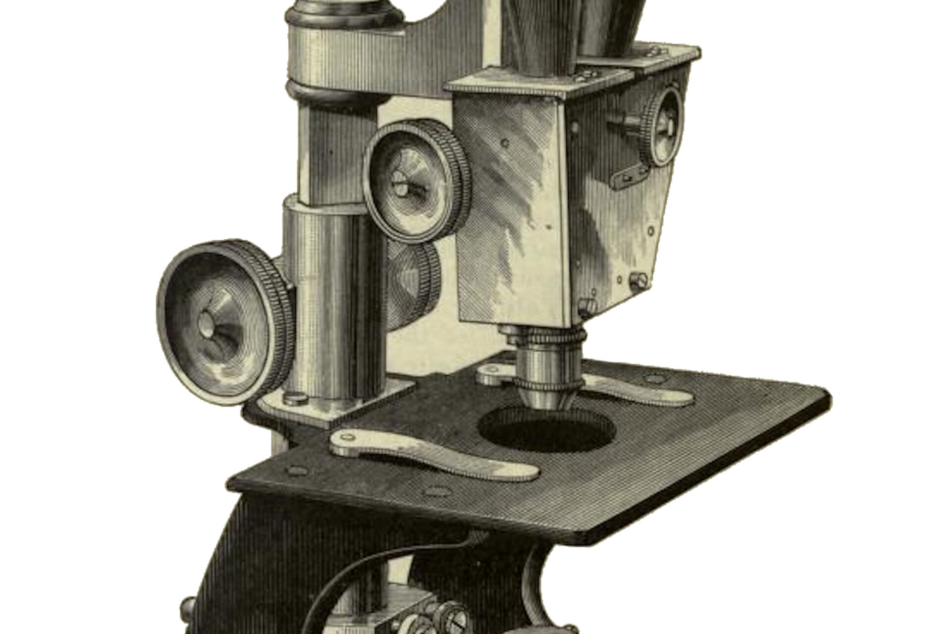
Gradually, standards were developed for microscope design and these were used by many manufacturers of optical instruments in the early days of industrial mass production (refer to figure 1). Advances in microscope design made possible groundbreaking discoveries in the fields of botany, histology, cytology, bacteriology, and medicine, such as the medical advances by Robert Koch [1] and Rudolf Virchow [2]. Suitable methods for fixing, embedding, and sectioning specimens, e.g. using a microtome, as well as specialized stains and preservatives were developed at the same time.

A monk designed the first stereoscopic microscope
A genuine stereo microscope allows each eye of the observer to observe the sample through a separate, dedicated light path, similar to having 2 single eyepiece microscopes combined into one. In parallel to the development of monocular telescopes and microscopes, work was already being done to design instruments for both eyes in the 17th century. Inspired by the description of a binocular microscope from the Capuchin monk Antonius Maria de Rheita dating from 1645, his fellow monk, Chérubin d’Orléans, applied in 1677 the familiar principle of the binocular telescope to the design of a microscope that would permit tiny objects to be viewed with both eyes at the same time (refer to figure 2). His goal was not a 3-dimensional image or depth perception; he believed that image quality could be improved by viewing objects with both eyes at the same time. The principle of stereoscopic vision was not yet known, as it was first described by the English physicist Charles Wheatstone in the year 1832.

In 1853, John Leonhard Riddel, a chemistry professor and postmaster in New Orleans, presented a binocular microscope with a single objective and a prism system (refer to figure 3).The prisms were arranged so that the right eye only received light from the right half of the objective and vice versa. The image was three-dimensional, but confusing because the relief appeared reversed (pseudoscopic).

Greenough and Cycloptic principles
Binocular microscopes of the day featured a simple lens system and the same design as traditional compound microscopes. They only attained low magnifications and did not permit significant working distances. These dissecting microscopes, as they were then known, were used primarily in biology for dissection purposes; there were no technical applications for them at the time.
Around 1890, the American biologist and zoologist Horatio S. Greenough, the son of the well-known American sculptor of the same name (refer to figure 4), introduced a design principle which is still used today by all major manufacturers of optical instruments. Stereo microscopes based on the "Greenough principle" (refer to figure 5) deliver genuine stereoscopic images of very high quality.
In 1957 the American Optical Company introduced the modern stereo microscope design with a shared main objective and named it Cycloptic (refer to figure 6). Its modern aluminum housing contained two parallel beam paths and the main objective, as well as a five-step magnification changer. This stereo microscope type, which was based on the telescope or CMO (Common Main Objective) principle (refer to figure 7), was adopted in addition to the Greenough type by all manufacturers and used for modular, high-performance instruments. Two years later, another American company, Bausch & Lomb, presented its StereoZoom Greenough design with a groundbreaking innovation: a stepless magnification changer (zoom).

 in the beam paths ensure that the images are upright and correctly oriented.")
 principle.")
 system.")
Stereo microscopy today
Although the basic stereo microscope has been around for a very long time, it has more recently assumed an even more important role. Microscopes are often involved in the manufacture or development of many products used every day, especially those concerning high-tech applications like mobile devices. The same applies to watches, irrespective of whether they are luxury or economy models. Stereo microscopes are also used for medical technology products, e.g., artificial hearts, defibrillators, or stents.
However, the use of stereo microscopes is not confined to manufacturing. Also, they are often used for other applications, such as forensics concerning the gathering evidence at the microscopic level, e.g., small particles of material or textile fibers, for convicting criminals, as well as for research in the life sciences and material sciences.
References
- Robert Koch was a main pioneer in the fields of medical microbiology and bacteriology, discovered the bacteria anthrax bacillus, tuberculosis bacillus, and cholera bacillus, and was awarded the Nobel Prize for Medicine and Physiology in 1905.
- Rudolf Virchow was a main pioneer in the fields of cytopathology (study of disease at the cellular level), comparative pathology (study of diseases common to humans and animals), anthropology (study of humanity), and ethnology (anthropology focusing on cultures). He proposed the third dictum of cell theory: "Omnis cellula e cellula” (All cells come from cells).
- W.B. Carpenter, W.H. Dallinger, The Principles and Theory of Vision with the Compound Microscope, Ch. II in The Microscope and Its Revelations, 8th Ed. (P. Blakiston's Son & Co, Philadelphia, 1901) fig. 71, p. 96.
- W.B. Carpenter, W.H. Dallinger, The History and Development of the Microscope, Ch. III in The Microscope and Its Revelations, 8th Ed. (P. Blakiston's Son & Co, Philadelphia, 1901) fig. 98, p. 131.
- Frances Benjamin Johnston Photograph Collection, USA Library of Congress, Prints & Photographs Division, Reproduction number LC-DIG-ppmsc-04904.
Related Articles
-

Epi-Illumination Fluorescence and Reflection-Contrast Microscopy
This article discusses the development of epi-illumination and reflection contrast for fluorescence…
Nov 02, 2023Read article -
 illuminated with wide-band UV excitation. Note the tissue structure with blue autofluorescence. Right: Same tissue and same immunostaining with FITC label illuminated with epi-illumination using narrow")
Milestones in Incident Light Fluorescence Microscopy
Since the middle of the last century, fluorescence microscopy developed into a bio scientific tool…
Mar 06, 2017Read article -
")
A Brief History of Light Microscopy
The history of microscopy begins in the Middle Ages. As far back as the 11th century, plano-convex…
Sep 08, 2015Read article
