
Science Lab
Science Lab
The knowledge portal of Leica Microsystems offers scientific research and teaching material on the subjects of microscopy. The content is designed to support beginners, experienced practitioners and scientists alike in their everyday work and experiments. Explore interactive tutorials and application notes, discover the basics of microscopy as well as high-end technologies – become part of the Science Lab community and share your expertise!
Filter articles
Tags
Story Type
Products
Loading...

Potential of Multiplex Confocal Imaging for Cancer Research and Immunology
Explore the new frontiers of multi-color fluorescent imaging: from image acquisition to analysis
Loading...
. Courtesy: Thomas Mathivet, PhD")
Windows on Neurovascular Pathologies
Discover how innate immunity can sustain deleterious effects following neurovascular pathologies and the technological developments enabling longitudinal studies into these events.
Loading...
 and labelled with MitoTracker Green.")
The Power of Reproducibility, Collaboration and New Imaging Technologies
In this webinar you willl learn what impacts reproducibility in microscopy, what resources and initiatives there are to improve education and rigor and reproducibility in microscopy and how…
Loading...
Tracking Single Cells Using Deep Learning
AI-based solutions continue to gain ground in the field of microscopy. From automated object classification to virtual staining, machine and deep learning technologies are powering scientific…
Loading...

Learning the Cellular Architecture from its Optical Properties
In the last 3 years, microscopists have started to use "AI based" solutions for a wide range of applications, including image acquisition optimization (smart microscopy), object classification, image…
Loading...

A Quality Metric for the Systematic Evaluation of Clearing Protocols
3D multicellular spheroids are of interest for studying tumor behavior and evaluating the response of pharmacologically active agents, because they mimic the in vivo tumor environment better than…
Loading...
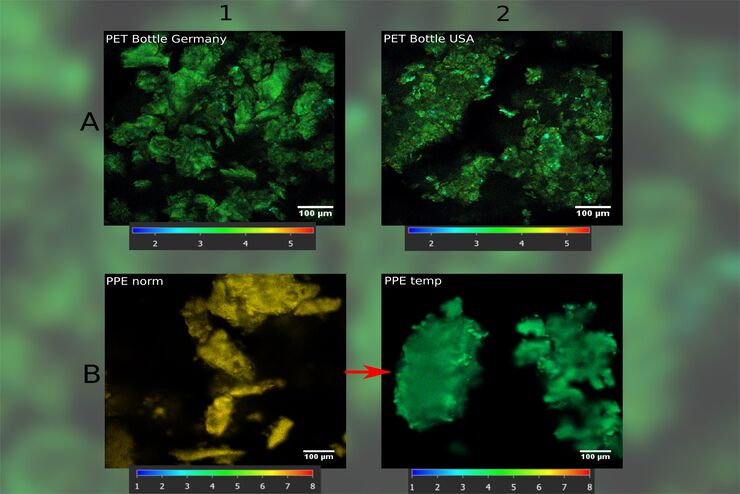
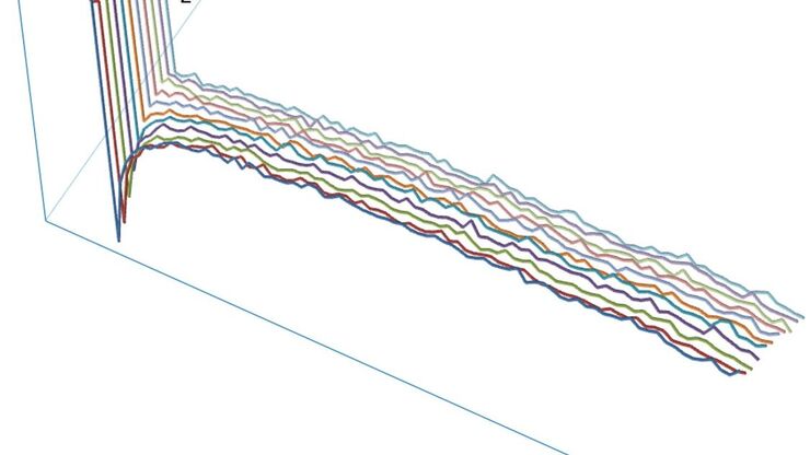
How FLIM Microscopy Helps to Detect Microplastic Pollution
The use of autofluorescence in biological samples is a widely used method to gain detailed knowledge about systems or organisms. This property is not only found in biological systems, but also…
Loading...

Development of Fluorescence Lifetime Imaging Microscopy (FLIM) and its Relevance for Functional Imaging
Prof. Ammasi Periasamy, Director, Keck Center for Cellular Imaging, University of Virginia, was interviewed by Dr. Giulia Ossato, Product Manager functional imaging, during Leica Microsystems Meets…
Loading...
Step by Step Guide for FRAP Experiments
Fluorescence Recovery After Photobleaching (FRAP) has been considered the most widely applied method for observing translational diffusion processes of macromolecules. The resulting information can be…